. 2012 Apr;33(12):3401-10.
doi: 10.1016/j.biomaterials.2012.01.012. Epub 2012 Feb 3.

A rat decellularized small bowel scaffold that preserves villus-crypt architecture for intestinal regeneration

Affiliations

A rat decellularized small bowel scaffold that preserves villus-crypt architecture for intestinal regeneration

Giorgia Totonelli et al. Biomaterials. 2012 Apr.

Abstract

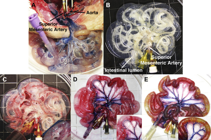
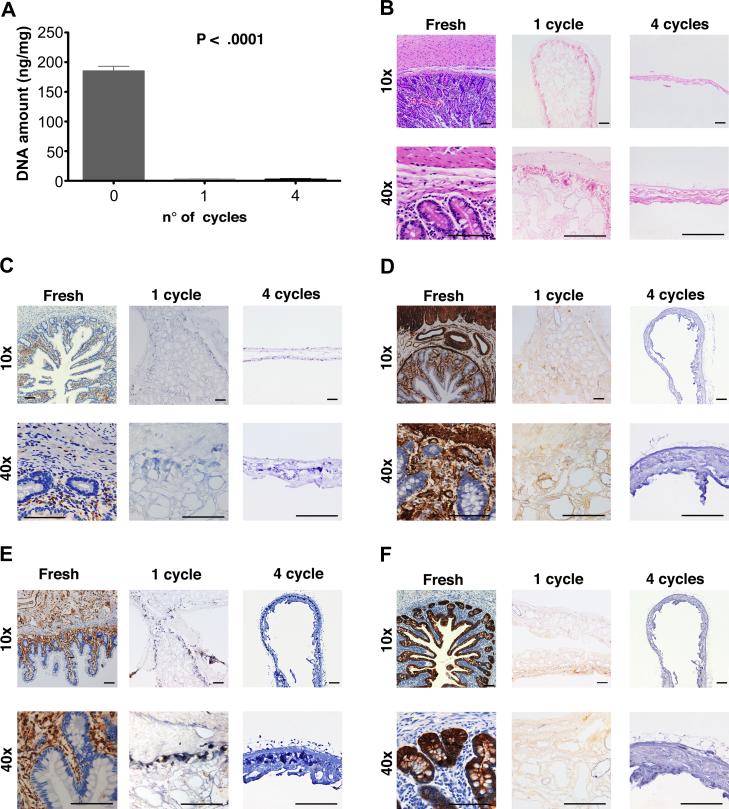
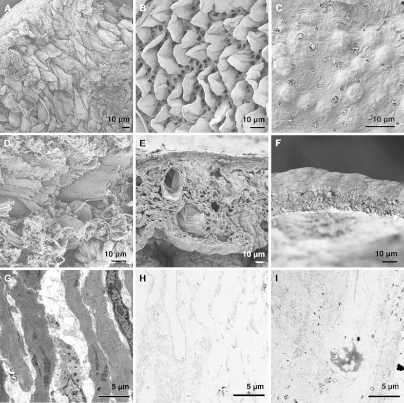
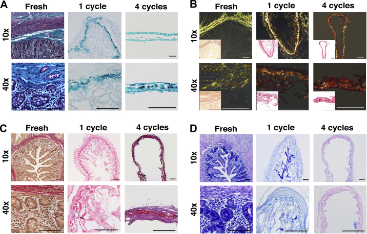
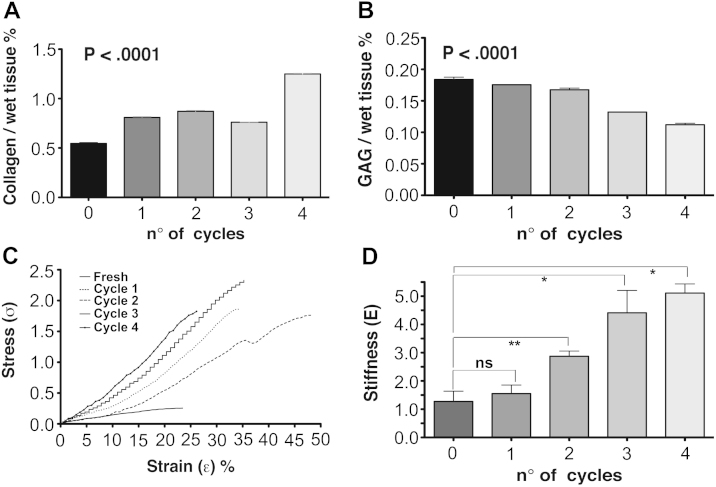
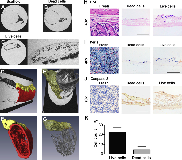
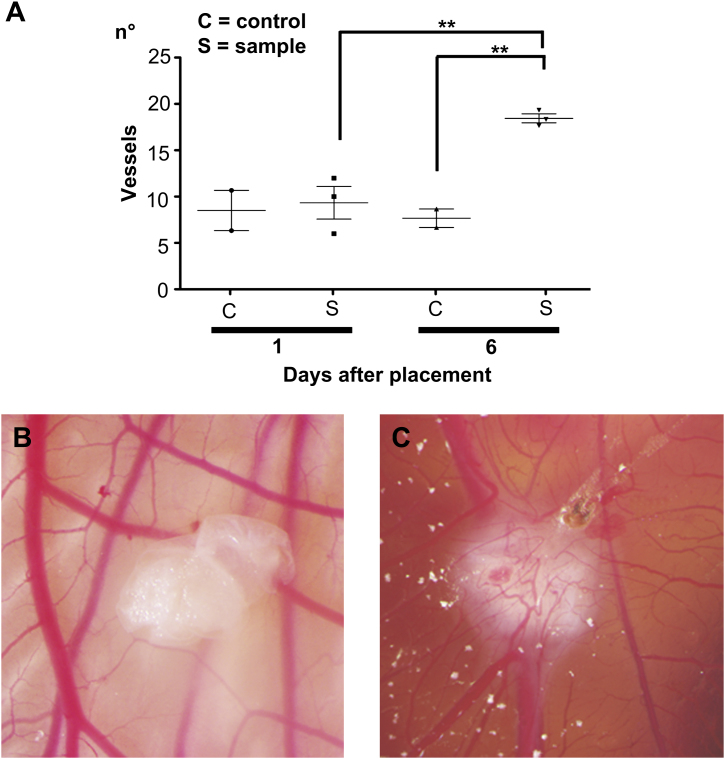
Management of intestinal failure remains a clinical challenge and total parenteral nutrition, intestinal elongation and/or transplantation are partial solutions. In this study, using a detergent-enzymatic treatment (DET), we optimize in rats a new protocol that creates a natural intestinal scaffold, as a base for developing functional intestinal tissue. After 1 cycle of DET, histological examination and SEM and TEM analyses showed removal of cellular elements with preservation of the native architecture and connective tissue components. Maintenance of biomechanical, adhesion and angiogenic properties were also demonstrated strengthen the idea that matrices obtained using DET may represent a valid support for intestinal regeneration.


Figures

Fig. 1
Fig. 1
Decellularization of rat small intestine with detergent-enzymatic treatment. Macroscopic images prior (A) and following (B) one cycle of decellularization. Perfusion with Rosso Ponceau (C), Trypan blue (D) and Rhodamine green (E) dyes displays the patency and distribution of the arterial (C) and venous (D) capillary beds in contrast with the lumen (E) proving absence of leakage.
Fig. 2
Fig. 2
DNA quantification shows complete removal of DNA following 1 cycle of detergent-enzymatic treatment (P < 0.001) with no significant differences in DNA with additional cycles (A). H&E staining confirms the absence of nuclei and demonstrates preservation of structure following 1 cycle of treatment compared to fresh tissue and the loss of this upon prolonged decellularization as in cycle 4 (B). The lack of immunogenicity is confirmed with immunostaining for MHC-II (C). Immunostaining for SMA (D), Vimentin (E) and MNF 116 (F) confirms the absence of both mesoderm-derived and epithelial tissue markers with 1 cycle of treatment. The architecture of mucosa, submucosa and muscularis propria is lost with further cycles; H&E: hematoxylin and eosin, MHC-II: major histocompatibility complex II, SMA: smooth muscle actin, MNF 116: pan-cytokeratin.
Fig. 3
Fig. 3
SEM and TEM of fresh intestine (A) and following 1 (B) and 4 (C) cycles of decellularization. While at 1 cycle the crypt/villus structure was completely preserved (B), this was completely lost at 4 cycles (C). SEM: scanning electron microscopy, TEM: transmission electron microscopy.
Fig. 4
Fig. 4
Characterization of the scaffold demonstrates preservation of structure, components of the ECM and removal of cellular elements. Masson’s Trichrome (A) and Picrosirius Red (B) staining confirm the maintenance of the connective tissue and collagen component of ECM. EVG staining (C) confirms the preservation of the elastin component around the blood vessels (black arrow) and Alcian Blue staining (D) the preservation of glycosaminoglycans; ECM: extracellular matrix, EVG: Elastic van Gieson.
Fig. 5
Fig. 5
Collagen and GAG content of native tissue and decellularized samples at different cycles of DET (n ≥ 3 samples for all measures). Collagen content is increased in the acellular matrix (P < 0.0001; A) whilst GAG amount progressively decreases (P < 0.0001; B). Mechanical characterization of the acellular matrix: stress–strain curves show the tensile strength increasing with the number of cycles (C). No significant difference is observed in term of stiffness between native tissue and acellular matrix. (n ≥ 5 samples for all measures) (D); GAG: glycosaminoglycans.
Fig. 6
Fig. 6
Magnetic resonance imaging as a viability test (A–K). Imaging of the acellular matrix shows its cylindrical structure with mesentery on the side (A). Matrix seeded with apoptotic AFSC shows partial repopulation and dissimilar distribution of the cells on the luminal surface (B). Scaffold seeded with live cells shows uniform repopulation of the lumen with cells distributed on the villi (both axial and longitudinal section) (C). Three-dimensional reconstruction demonstrates the matrix (red) overlying the cell layer (gray) and mesentery on the side (yellow) (D–G).H&E (H), Prussian blue (I), and Cleaved Caspase 3 (J) stains of scaffold seeded with both live and dead cells. Scale bars, 100 μm. Counting of cells seeded onto the matrix: number of live cells is significantly higher than dead cells (P = 0.01)(n = 5 for each measure) (K); AFSC: amniotic fluid stem cells, H&E: hematoxylin and eosin. (For interpretation of the references to color in this figure legend, the reader is referred to the web version of this article.)
Fig. 7
Fig. 7
Pro-angiogenic properties of intestinal acellular matrix in vivo (A–C). Macroscopic quantification of converging vessels was blindly made for both intestinal decellularized samples and polyester membrane used as negative control (A). On day 6 after implantation, the number of vessels converging towards the intestinal matrices is significantly increased in comparison to the same samples at day 1 (P < 0.01) and to the polyester membrane that was used as a negative control (P < 0.05). Example of CAM at 1 day after implantation of intestinal acellular matrix: the sample of decellularized tissue is adherent to the CAM and starts to be surrounded by allantoic vessels (B). After 6 days of implantation, intestinal matrices are completely enveloped by the newly formed vessels, organized in a network (C).
Video S1
Video S1
This movie shows high-resolution magnetic resonance (MRI) images of cells labeled with micron sized iron oxide particles seeded onto a natural small intestine scaffold. The movie starts transversing through short axis MRI images along the major axis of the intestine. A few seconds after the start, the volume rendering of segmented cells (brown, threshold of hypointensities), the intestine membrane (red) and the mesentery (yellow) is activated. (MPEG; 27 MB).

References

    1. Lloyd D.A., Vega R., Bassett P., Forbes A., Gabe S.M. Survival and dependence on home parenteral nutrition: experience over a 25-year period in a UK referral centre. Aliment Pharmacol Ther. 2006;24:1231–1240. - PubMed
    1. Goulet O., Ruemmele F., Lacaille F., Colomb V. Irreversible intestinal failure. J Pediatr Gastroenterol Nutr. 2004;38:250–269. - PubMed
    1. Bianchi A. Intestinal loop lengthening–a technique for increasing small intestinal length. J Pediatr Surg. 1980;15:145–151. - PubMed
    1. Vianna R.M., Mangus R.S. Present prospects and future perspectives of intestinal and multivisceral transplantation. Curr Opin Clin Nutr Metab Care. 2009;12:281–286. - PubMed
    1. Kemp P. History of regenerative medicine: looking backwards to move forwards. Regen Med. 2006;1:653–669. - PubMed

Publication types