

Chronic pneumonia in calves after experimental infection with Mycoplasma bovis strain 1067: characterization of lung pathology, persistence of variable surface protein antigens and local immune response
- PMID: 22305416
- PMCID: PMC3287114
- DOI: 10.1186/1751-0147-54-9
Chronic pneumonia in calves after experimental infection with Mycoplasma bovis strain 1067: characterization of lung pathology, persistence of variable surface protein antigens and local immune response
Abstract
Background: Mycoplasma bovis is associated with pneumonia in calves characterized by the development of chronic caseonecrotic lesions with the agent persisting within the lesion. The purposes of this study were to characterize the morphology of lung lesions, examine the presence of M. bovis variable surface protein (Vsp) antigens and study the local immune responses in calves after infection with M. bovis strain 1067.
Methods: Lung tissue samples from eight calves euthanased three weeks after experimental infection with M. bovis were examined by bacteriology and pathology. Lung lesions were evaluated by immunohistochemical (IHC) staining for wide spectrum cytokeratin and for M. bovis Vsp antigens and pMB67 antigen. IHC identification and quantitative evaluation of CD4+ and CD8+ T lymphocytes and immunoglobulin (IgG1, IgG2, IgM, IgA)-containing plasma cells was performed. Additionally, expression of major histocompatibility complex class II (MHC class II) was studied by IHC.
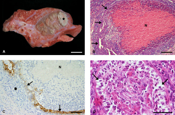
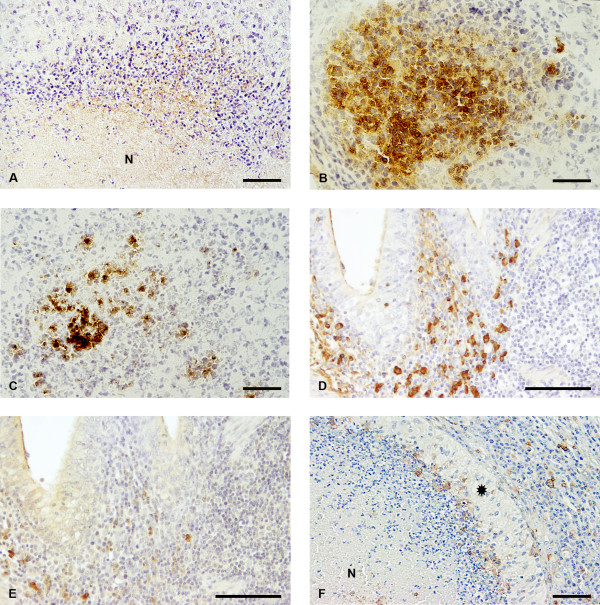
Results: Suppurative pneumonic lesions were found in all calves. In two calves with caseonecrotic pneumonia, necrotic foci were surrounded by epithelial cells resembling bronchial or bronchiolar epithelium. In all calves, M. bovis Vsp antigens were constantly present in the cytoplasm of macrophages and were also present extracellularly at the periphery of necrotic foci. There was a considerable increase in numbers of IgG1- and IgG2-positive plasma cells among which IgG1-containing plasma cells clearly predominated. Statistical evaluation of the numbers of CD4+ and CD8+ T cells, however, did not reveal statistically significant differences between inoculated and control calves. In M. bovis infected calves, hyperplasia of bronchus-associated lymphoid tissue (BALT) was characterized by strong MHC class II expression of lymphoid cells, but only few of the macrophages demarcating the caseonecrotic foci were positive for MHC class II.
Conclusions: The results from this study show that infection of calves with M. bovis results in various lung lesions including caseonecrotic pneumonia originating from bronchioli and bronchi. There is long-term persistence of M. bovis as demonstrated by bacteriology and immunohistochemistry for M. bovis antigens, i.e. Vsp antigens and pMB67. The persistence of the pathogen and its ability to evade the specific immune response may in part result from local downregulation of antigen presenting mechanisms and an ineffective humoral immune response with prevalence of IgG1 antibodies that, compared to IgG2 antibodies, are poor opsonins.
Figures
Similar articles
-
Histopathological findings, phenotyping of inflammatory cells, and expression of markers of nitritative injury in joint tissue samples from calves after vaccination and intraarticular challenge with Mycoplasma bovis strain 1067.Acta Vet Scand. 2014 Aug 19;56(1):45. doi: 10.1186/s13028-014-0045-3. Acta Vet Scand. 2014. PMID: 25162202 Free PMC article.
-
Long-term survival of Mycoplasma bovis in necrotic lesions and in phagocytic cells as demonstrated by transmission and immunogold electron microscopy in lung tissue from experimentally infected calves.Vet Microbiol. 2013 Mar 23;162(2-4):949-953. doi: 10.1016/j.vetmic.2012.11.039. Epub 2012 Dec 7. Vet Microbiol. 2013. PMID: 23294861
-
Effects of inflammatory stimuli on the development of Mycoplasma bovis pneumonia in experimentally challenged calves.Vet Microbiol. 2024 Oct;297:110203. doi: 10.1016/j.vetmic.2024.110203. Epub 2024 Jul 30. Vet Microbiol. 2024. PMID: 39089141
-
Proteomics analysis and its role in elucidation of functionally significant proteins in Mycoplasma bovis.Microb Pathog. 2017 Oct;111:50-59. doi: 10.1016/j.micpath.2017.08.024. Epub 2017 Aug 18. Microb Pathog. 2017. PMID: 28826762 Review.
-
Mycoplasma bovis in respiratory disease of feedlot cattle.Vet Clin North Am Food Anim Pract. 2010 Jul;26(2):365-79. doi: 10.1016/j.cvfa.2010.03.003. Epub 2010 May 14. Vet Clin North Am Food Anim Pract. 2010. PMID: 20619190 Review.
Cited by
-
Unveiling the stealthy tactics: mycoplasma's immune evasion strategies.Front Cell Infect Microbiol. 2023 Aug 31;13:1247182. doi: 10.3389/fcimb.2023.1247182. eCollection 2023. Front Cell Infect Microbiol. 2023. PMID: 37719671 Free PMC article. Review.
-
Identification and subtyping of clinically relevant human and ruminant mycoplasmas by use of matrix-assisted laser desorption ionization-time of flight mass spectrometry.J Clin Microbiol. 2013 Oct;51(10):3314-23. doi: 10.1128/JCM.01573-13. Epub 2013 Jul 31. J Clin Microbiol. 2013. PMID: 23903545 Free PMC article.
-
Molecular cloning and characterization of a surface-localized adhesion protein in Mycoplasma bovis Hubei-1 strain.PLoS One. 2013 Jul 23;8(7):e69644. doi: 10.1371/journal.pone.0069644. Print 2013. PLoS One. 2013. PMID: 23936063 Free PMC article.
-
Mycoplasma bovis and viral agents associated with the development of bovine respiratory disease in adult dairy cows.Transbound Emerg Dis. 2020 Jul;67 Suppl 2(Suppl 2):82-93. doi: 10.1111/tbed.13223. Epub 2019 Jun 24. Transbound Emerg Dis. 2020. PMID: 31232526 Free PMC article.
-
Mycoplasma bovis is associated with Mannheimia haemolytica during acute bovine respiratory disease in feedlot cattle.Front Microbiol. 2022 Aug 1;13:946792. doi: 10.3389/fmicb.2022.946792. eCollection 2022. Front Microbiol. 2022. PMID: 35979489 Free PMC article.
References
MeSH terms
Substances
LinkOut - more resources
Full Text Sources
Research Materials
Miscellaneous
