. 2012 Feb 3;10(2):210-7.
doi: 10.1016/j.stem.2012.01.004.

Decoupling of tumor-initiating activity from stable immunophenotype in HoxA9-Meis1-driven AML

Affiliations

Decoupling of tumor-initiating activity from stable immunophenotype in HoxA9-Meis1-driven AML

Kenneth D Gibbs Jr et al. Cell Stem Cell. .

Erratum in

  • Cell Stem Cell. 2012 Apr 6;10(4):480

Abstract

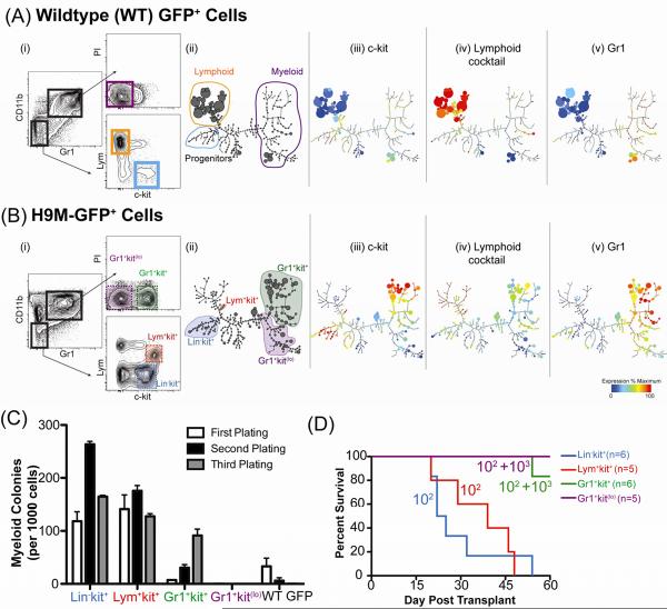
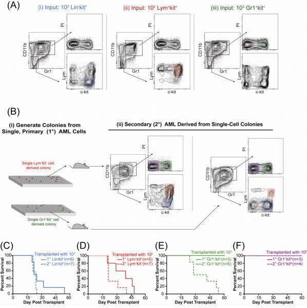
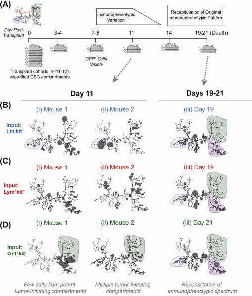
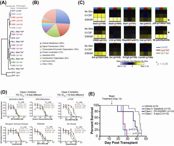
Increasing evidence suggests tumors are maintained by cancer stem cells; however, their nature remains controversial. In a HoxA9-Meis1 (H9M) model of acute myeloid leukemia (AML), we found that tumor-initiating activity existed in three, immunophenotypically distinct compartments, corresponding to disparate lineages on the normal hematopoietic hierarchy--stem/progenitor cells (Lin(-)kit(+)) and committed progenitors of the myeloid (Gr1(+)kit(+)) and lymphoid lineages (Lym(+)kit(+)). These distinct tumor-initiating cells (TICs) clonally recapitulated the immunophenotypic spectrum of the original tumor in vivo (including cells with a less-differentiated immunophenotype) and shared signaling networks, such that in vivo pharmacologic targeting of conserved TIC survival pathways (DNA methyltransferase and MEK phosphorylation) significantly increased survival. Collectively, H9M AML is organized as an atypical hierarchy that defies the strict lineage marker boundaries and unidirectional differentiation of normal hematopoiesis. Moreover, this suggests that in certain malignancies tumor-initiation activity (or "cancer stemness") can represent a cellular state that exists independently of distinct immunophenotypic definition.


Figures

Figure 1
Figure 1. Multiple, Immunophenotypically Distinct Compartments Possess Tumor-Initiating Activity (TIA)
(A,B) Immunophenotypic comparison of bone marrow from mice transplanted with (A) GFP or (B) H9M-GFP transduced progenitors. (i) Flow cytometric analysis. “Lym” is a cocktail of B- and T-cell markers (B220, CD19, CD3ε, CD4, CD8a, and TCR-β). PI is propidium iodide. (ii) SPADE analysis of immunophenotypic progression. Size of each circle (metacell) indicates relative cell frequency. Metacell clusters were manually annotated based on (iii) c-kit, (iv) lymphoid cocktail, and (v) Gr1 expression. These parameters and CD11b used for tree drawing. (C) FACS-purified populations from H9M-GFP marrow were sorted and plated in methylcellulose with SCF, GM-CSF, IL-3, and IL-6 (S36GM). Colonies were scored after seven days, dissociated and replated. Numbers per 1000 cells plated (in triplicate ± SEM). Total WT-GFP cells shown as control. (D) Survival curves of mice lethally irradiated and transplanted with indicated numbers of cells (plus 105 helper marrow) from following compartments: Lin-kit+ (blue, 102 cells), Lym+kit+ (red, 102 cells), Gr1+kit+ (green, 102 or 103 cells), or Gr1+kitlo (purple, 102 or 103 cells). See also Figures S1–2.
Figure 2
Figure 2. Enforced H9M Expression Permits Surface Marker Plasticity in TIC Resulting in a Dynamic Tumorigenic Hierarchy
(A) Flow cytometry of representative marrow from secondary mice transplanted with indicated cell number from specified compartment. Shaded region indicates the population transplanted. (B) (i) Single cells from primary AML were expanded into colonies and after one week transplanted into secondary mice. (ii) Flow cytometric analysis of the GFP+ marrow compartment of secondary mice transplanted with single-cell derived Lym+kit+ colony (top) or Gr1+kit+ colony (bottom). (C–F) Survival curves from mice transplanted with 100 primary or secondary (C) Lin-kit+, (D) Lym+kit+, (E) Gr1+kit+, or (F) Gr1+kitlo cells. See also Figure S3.
Figure 3
Figure 3. In Vivo Tumorigenic Differentiation Does Not Proceed Along a Strictly Defined Pathway
(A) Experimental scheme. Defined numbers of each TIC in primary AML were transplanted into cohorts of secondary recipients. Animals were sacrificed every 3–4 four days thereafter to assess for frequency and immunophenotype of progeny. (B–D). SPADE analysis of bone marrow of individual mice transplanted with (B) 500 Lin-kit+ cells, (C) 500 Lym+kit+ cells, or (D) 2500 Gr1+kit+ cells on (i, ii) day 11 post transplant or (iii) 19–21 days post transplant. Shaded regions in panels i and ii are the compartment transplanted, and in panel iii are compartments present in primary leukemia. See also Figure S4.
Figure 4
Figure 4. Phenotypically Distinct TICs Have Conserved Signaling and Survival Networks & Targeting Conserved Nodes in vivo Increases Survival
(A) Hierarchical clustering of phenotypic compartments from primary H9M AML (H9M: Lin-kit+, Lym+kit+, Gr1+kit+, Gr1+kitlo), populations from MLL-AF9 AML with high or low TIA (Mac1+kit+ or Mac1+kitlo, respectively), and analogous populations from GFP marrow (Lin-kit+, Lym+, and Gr1+). Each line represents a unique biological replicate. (B) GO analysis of genes upregulated in primary H9M TICs (Lin-kit+ and Lym+kit+) relative to the Gr1+kit+ compartment based on biological process annotations. See also Figure S5 & Table S1. (C) Heatmap of the phospho-response in TICs (colored bar indicates compartment) isolated from secondary AML, measured by mass cytometry. ANOVA was used to test for statistically significant differences in fold-change values between TIC in all mice tested (n=3). Asterisks indicate statistically significant differences (p<0.05). See also Figure S6. (D) Defined numbers of cells from each TIC (Lin-kit+, blue; Lym+kit+, red; Gr1+kit+, green) were sorted into liquid medium with S36GM and concentrations of inhibitors for the pathways indicated above the plots. Plots represent numbers after 7 days measured quadruplicate ± SEM. (E) Survival curves of secondary mice transplanted with 104 total GFP+ primary AML marrow cells treated with dasatinib, PD-0325901, and 5-aza-2'-deoxycytidine. Survival differences between mice treated vehicle and PD-0325901 (p=0.01, Log-Rank Test) or 5-aza-2'-deoxycytidine (p<0.0001, Log-Rank Test) are statistically significant.

Comment in

References

    1. Abramovich C, Humphries RK. Hox regulation of normal and leukemic hematopoietic stem cells. Current opinion in hematology. 2005;12:210–216. - PubMed
    1. Anderson K, Lutz C, van Delft FW, Bateman CM, Guo Y, Colman SM, Kempski H, Moorman AV, Titley I, Swansbury J, et al. Genetic variegation of clonal architecture and propagating cells in leukaemia. Nature. 2011;469:356–361. - PubMed
    1. Bendall SC, Simonds EF, Qiu P, Amir el AD, Krutzik PO, Finck R, Bruggner RV, Melamed R, Trejo A, Ornatsky OI, et al. Single-cell mass cytometry of differential immune and drug responses across a human hematopoietic continuum. Science. 2011;332:687–696. - PMC - PubMed
    1. Bonnet D, Dick JE. Human acute myeloid leukemia is organized as a hierarchy that originates from a primitive hematopoietic cell. Nature medicine. 1997;3:730–737. - PubMed
    1. Clarke MF, Dick JE, Dirks PB, Eaves CJ, Jamieson CH, Jones DL, Visvader J, Weissman IL, Wahl GM. Cancer Stem Cells--Perspectives on Current Status and Future Directions: AACR Workshop on Cancer Stem Cells. Cancer research. 2006;66:9339–9344. - PubMed

Publication types

MeSH terms

Substances