

Molecular mechanisms underlying chemical liver injury
- PMID: 22306029
- PMCID: PMC3704158
- DOI: 10.1017/S1462399411002110
Molecular mechanisms underlying chemical liver injury
Abstract
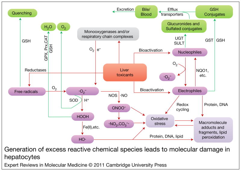
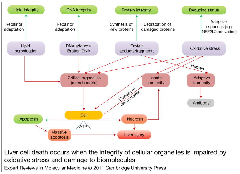
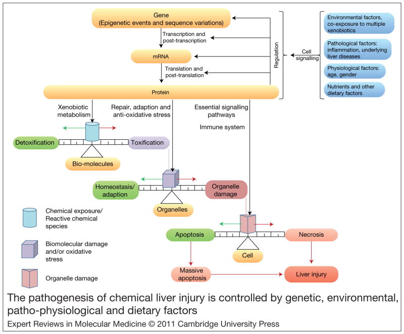
The liver is necessary for survival. Its strategic localisation, blood flow and prominent role in the metabolism of xenobiotics render this organ particularly susceptible to injury by chemicals to which we are ubiquitously exposed. The pathogenesis of most chemical-induced liver injuries is initiated by the metabolic conversion of chemicals into reactive intermediate species, such as electrophilic compounds or free radicals, which can potentially alter the structure and function of cellular macromolecules. Many reactive intermediate species can produce oxidative stress, which can be equally detrimental to the cell. When protective defences are overwhelmed by excess toxicant insult, the effects of reactive intermediate species lead to deregulation of cell signalling pathways and dysfunction of biomolecules, leading to failure of target organelles and eventual cell death. A myriad of genetic factors determine the susceptibility of specific individuals to chemical-induced liver injury. Environmental factors, lifestyle choices and pre-existing pathological conditions also have roles in the pathogenesis of chemical liver injury. Research aimed at elucidating the molecular mechanism of the pathogenesis of chemical-induced liver diseases is fundamental for preventing or devising new modalities of treatment for liver injury by chemicals.
Figures
References
-
- Nemeth E, Baird AW, O’Farrelly C. Microanatomy of the liver immune system. Seminars in Immunopathology. 2009;31:333–343. - PubMed
-
- Dienes HP, Drebber U. Pathology of immune-mediated liver injury. Digestive Diseases. 2010;28:57–62. - PubMed
-
- Misdraji J. Embryology, anatomy, histology, and developmental anomalies of the liver. In: Sleisenger MH, Feldman M, et al., editors. Sleisenger and Fordtran’s Gastrointestinal and Liver Disease: Pathophysiology, Diagnosis, Management. 9. Saunders/Elsevier; Philadelphia, PA: 2010. pp. 1201–1206.
-
- Carithers RL, Jr, Mcclain CJ. Alcoholic liver disease. In: Sleisenger MH, Feldman M, et al., editors. Sleisenger and Fordtran’s Gastrointestinal and Liver Disease: Pathophysiology, Diagnosis, Management. 9. Saunders/Elsevier; Philadelphia, PA: 2010. pp. 1383–1400.
-
- Jungermann K, Kietzmann T. Zonation of parenchymal and nonparenchymal metabolism in liver. Annual Review of Nutrition. 1996;16:179–203. - PubMed
Further reading, resources and contacts
-
- Gregus Z. Mechanism of toxicity. In: Casarett LJ, Doull J, et al., editors. Casarett and Doull’s Toxicology: The Basic Science of Poisons. 7. McGraw-Hill; New York: 2008. pp. 45–106. This book chapter provides the most comprehensive and detailed information on the molecular and cellular mechanism of toxicity by xenobiotics.
-
- Kaplowitz N. Biochemical and cellular mechanisms of toxic liver injury. Seminars in Liver Disease. 2002;22:137–144. This review focuses on the mechanisms of different forms of cell death. - PubMed
-
- Abboud G, Kaplowitz N. Drug-induced liver injury. Drug Safety. 2007;30:277–294. This review provides practical information on the diagnosis and management of patients with suspected drug-related liver disease. Pathogenesis is also discussed briefly. - PubMed
-
- Testa B, Kramer SD. The biochemistry of drug metabolism – an introduction: parts 2–5. Redox, hydrolysis, conjugation reactions and their enzymes, metabolism and bioactivity. Chemistry and Biodiversity. 2007–2009;4456:257–405. 2031–2122, 2171–2336, 591–684. - PubMed
-
- Parkinson A, Ogilvie BW. Biotransformation of xenobiotics. In: Casarett LJ, Doull J, et al., editors. Casarett and Doull’s Toxicology: The Basic Science of Poisons. 7. McGraw-Hill; New York: 2008. pp. 161–304. The reviews by Test and Kramer and the book chapter by Parkinson and Ogilvie demonstrate comprehensive and detailed information on detoxification and bio-activation of xenobiotics.
Publication types
MeSH terms
Substances
Grants and funding
LinkOut - more resources
Full Text Sources
Other Literature Sources
Medical
