Review
. 2012 Jun;23(4):450-7.
doi: 10.1016/j.semcdb.2012.01.010. Epub 2012 Jan 28.

Notch signaling in human development and disease

Affiliations
Review

Notch signaling in human development and disease

Andrea L Penton et al. Semin Cell Dev Biol. 2012 Jun.

Abstract

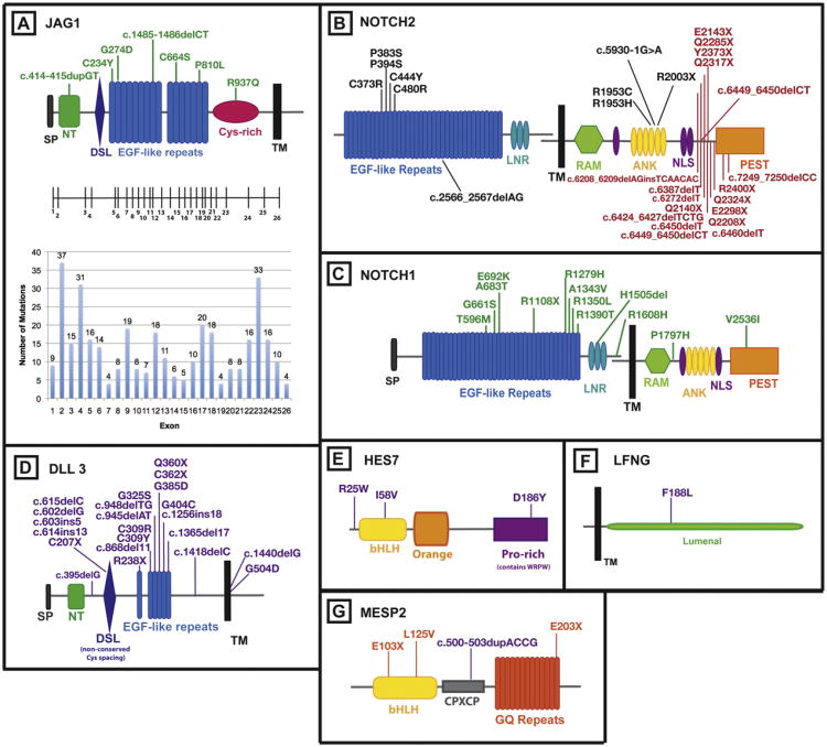
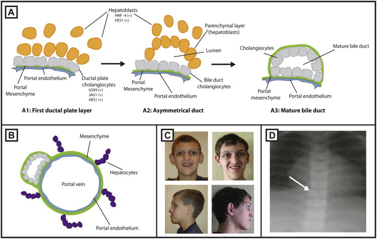
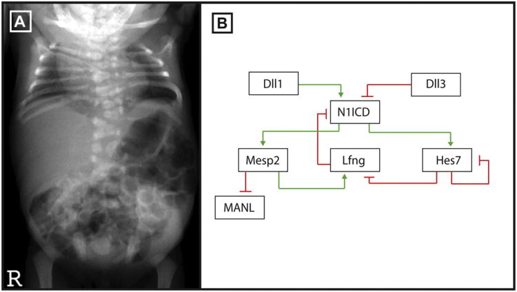
Mutations in Notch signaling pathway members cause developmental phenotypes that affect the liver, skeleton, heart, eye, face, kidney, and vasculature. Notch associated disorders include the autosomal dominant, multi-system, Alagille syndrome caused by mutations in both a ligand (Jagged1 (JAG1)) and receptor (NOTCH2) and autosomal recessive spondylocostal dysostosis, caused by mutations in a ligand (Delta-like-3 (DLL3)), as well as several other members of the Notch signaling pathway. Mutations in NOTCH2 have also recently been connected to Hajdu-Cheney syndrome, a dominant disorder causing focal bone destruction, osteoporosis, craniofacial morphology and renal cysts. Mutations in the NOTCH1 receptor are associated with several types of cardiac disease and mutations in NOTCH3 cause the dominant adult onset disorder CADASIL (cerebral autosomal dominant arteriopathy with subcortical infarcts and leukoencephalopathy), a vascular disorder with onset in the 4th or 5th decades. Studies of these human disorders and their inheritance patterns and types of mutations reveal insights into the mechanisms of Notch signaling.


Figures

Fig. 1
Fig. 1
Notch signaling pathway genes with associated mutations in human disease. (A) The protein domains of JAG1 are shown, with the corresponding exon structure for the gene. The number of ALGS associated mutations identified in each exon is represented on the graph below the exon structure. Mutations in patients with isolated cardiac disease are indicated directly on the protein structure diagram (green). (B) The protein domains of NOTCH2 are shown. NOTCH2 mutations are found in Alagille syndrome (black) and Hajdu-Cheney syndrome (red). (C) The protein domains of NOTCH1 are shown. NOTCH1 mutations are found in patients with isolated cardiac defects (green). (D) The protein domains of DLL3 are shown. DLL3 mutations are found in patients with SCD (purple). (E) The HES7 protein domains are shown. HES7 mutations are found in patients with SCD (purple). (F) The LFNG protein domains are shown. A mutation in LFNG was identified in a patient with SCD (purple). (G) The protein domains of MESP2 are shown. Mutations in MESP2 are found in patients with SCD (purple) and STD (orange). [References for mutations are identified in the text.] ANK, ankryrin repeats; bHLH, basic-helix–loop–helix; CPXCP, CPXCP motif; Cys-rich, cysteine rich domain; DSL, Delta/Serrate/lag-2; EGF repeats, epidermal growth factor-like repeats; GQ repeats, glycine-glutamine repeats; LNR, LIN-12/Notch repeats; Lumenal, luminal domain; NLS, nuclear localization signal; NT, N-terminal domain; Orange, orange domain, PEST, proline/glutamic acid/serine/threonine rich domain; Pro-rich, proline rich domain; RAM, RBP-Jк-associated module; SP, signal peptide; TM, transmembrane domain; WRPW, tryptophan-arginine-proline-tryptophan domain.
Fig. 2
Fig. 2
Development of intrahepatic bile ducts. (A1) Cholangiocytes (gray) line up along the portal vein forming the ductal plate.Jag1, Hes1 and Sox9 are expressed by cells of this layer. (A2) Subsequently, hepatoblasts expressing Hes1 and HNF4 (yellow) come into contact with the ductal plate forming the outer (parenchymal) layer and move to create an asymmetric tubular structure. Unincorporated hepatoblasts and cholangiocytes are eliminated. (A3) The outer hepatoblasts differentiate into cholangiocytes to complete the formation of the duct. (B) A mature duct aligned along the portal vein, enclosed by a mesenchyme. Mature hepatocytes develop in close proximity to bile ducts (purple). (C) Representative facies of a patient with ALGS are shown in frontal and side view. (D) Anteroposterior X-ray of a patient with butterfly vertebrae.
Fig. 3
Fig. 3
Notch signaling is required for somitogenesis. (A) Anteroposterior X-ray of a patient with SCD due to a mutation in DLL3 courtesy of Dr. Peter Turnpenny. The thoracic and cervical spine have severe vertebral segmentation defects including hemivertebrae. (B) A negative feedback loop consisting of the Mesp2, Lfng, Hes7 and Dll3 genes regulate Notch signaling activity during somitogenesis.

References

    1. Ellisen LW, Bird J, West DC, Soreng AL, Reynolds TC, Smith SD, et al. TAN-1, the human homolog of the Drosophila notch gene, is broken by chromosomal translocations in T lymphoblastic neoplasms. Cell. 1991;66(4):649–61. - PubMed
    1. Joutel A, Corpechot C, Ducros A, Vahedi K, Chabriat H, Mouton P, et al. Notch3 mutations in CADASIL, a hereditary adult-onset condition causing stroke and dementia. Nature. 1996;383(6602):707–10. - PubMed
    1. Li L, Krantz ID, Deng Y, Genin A, Banta AB, Collins CC, et al. Alagille syndrome is caused by mutations in human Jagged1, which encodes a ligand for Notch1. Nat Genet. 1997;16(3):243–51. - PubMed
    1. Alagille D, Borde J, Habib EC, Thomassin N. Surgical attempts in atresia of the intrahepatic bile ducts with permeable extrahepatic bile duct. Study of 14 cases in children. Arch Fr Pediatr. 1969;26(1):51–71. - PubMed
    1. Riely CA, Cotlier E, Jensen PS, Klatskin G. Arteriohepatic dysplasia: a benign syndrome of intrahepatic cholestasis with multiple organ involvement. Ann Intern Med. 1979;91(4):520–7. - PubMed

Publication types

MeSH terms

Supplementary concepts