. 2012 May 4;418(3-4):226-36.
doi: 10.1016/j.jmb.2012.01.016. Epub 2012 Jan 27.

Small-angle X-ray scattering and single-molecule FRET spectroscopy produce highly divergent views of the low-denaturant unfolded state

Affiliations

Small-angle X-ray scattering and single-molecule FRET spectroscopy produce highly divergent views of the low-denaturant unfolded state

Tae Yeon Yoo et al. J Mol Biol. .

Abstract

The results of more than a dozen single-molecule Förster resonance energy transfer (smFRET) experiments suggest that chemically unfolded polypeptides invariably collapse from an expanded random coil to more compact dimensions as the denaturant concentration is reduced. In sharp contrast, small-angle X-ray scattering (SAXS) studies suggest that, at least for single-domain proteins at non-zero denaturant concentrations, such compaction may be rare. Here, we explore this discrepancy by studying protein L, a protein previously studied by SAXS (at 5 °C), which suggested fixed unfolded-state dimensions from 1.4 to 5 M guanidine hydrochloride (GuHCl), and by smFRET (at 25 °C), which suggested that, in contrast, the chain contracts by 15-30% over this same denaturant range. Repeating the earlier SAXS study under the same conditions employed in the smFRET studies, we observe little, if any, evidence that the unfolded state of protein L contracts as the concentration of GuHCl is reduced. For example, scattering profiles (and thus the shape and dimensions) collected within ∼4 ms after dilution to as low as 0.67 M GuHCl are effectively indistinguishable from those observed at equilibrium at higher denaturant. Our results thus argue that the disagreement between SAXS and smFRET is statistically significant and that the experimental evidence in favor of obligate polypeptide collapse at low denaturant cannot be considered conclusive yet.


Figures

Figure 1
Figure 1
The dimensions of unfolded protein L have been studied using both single-molecule Forster resonance energy transfer (smFRET) and small angle x-ray scattering (SAXS). The results of these studies, however, are highly discordant. Specifically, time-resolved (open squares) and equilibrium (filled squares) SAXS studies by Baker and co-workers, conducted at 2.5°C and 5°C respectively, suggest that the radius of gyration of the unfolded ensemble transiently populated prior to refolding in 1.4 M GuHCl is experimentally indistinguishable from the dimensions observed at equilibrium at higher denaturant (22). In contrast, smFRET studies by Sherman and Haran (8) and Eaton and co-workers (9) and suggest that, at least at 20°C, unfolded protein L contracts significantly upon being transferred from high denaturant to lower denaturant. Here we explore this discrepancy in greater depth by performing more detailed SAXS studies under solution conditions mimicking those employed in the smFRET studies at both 5°C and 20°C. Of note, while the two smFRET data sets differ quantitatively -perhaps due to differences in the parameters used in the data analysis or due to a one residue-difference between the two constructs employed [see analysis in (42)], both argue in favor of significant collapse at low denaturant concentrations.
Figure 2
Figure 2
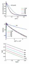
(top) The raw scattering profiles (I(0)-normalized scattering intensity versus q) of protein L collected at equilibrium at high denaturant are effectively indistinguishable over the range 3.5 to 7 M GuHCl, suggesting that the dimensions and shape of the unfolded protein do not change significantly over this range. At 1 M GuHCl, in contrast, the protein folds into its compact native state, dramatically altering its scattering. The data presented here is our APS data set; see Fig. S1 for the equivalent, independently collected CHESS results. CHESS data acquired at both 5°C and 20°C are also indistinguishable from one another (see Fig. S2), ruling out temperature dependent effects. (middle) Guinier representations of the scattering data likewise suggest that equilibrium unfolded protein L undergoes little if any change in dimensions down to at least 3.5 M GuHCl. (bottom) Finally, analysis of the fractal dimensions (30, 31) of unfolded protein L likewise suggests that its unfolded state remains expanded from 7 to 3.5 M GuHCl, as the slopes of log(I) versus log (q) plots (corresponding to the fractal dimension, Dm) collected under these conditions all fall between 1.52±0.01 to 1.62±0.01. The observed slopes approximate the 1.7 expected for an excluded volume random coil and fall far below the value of 3 expected for a compact globule.
Figure 3
Figure 3
A fit of the equilibrium SAXS data (APS data at 22°C; see Fig. S4 for the equivalent CHESS data set) to two-state equilibrium unfolding models does not produce any statistically significant evidence of a sloping unfolded baseline. Specifically, a model lacking baseline slope (solid line) fits the data well (R2 = 0.972), and the addition of a sloped unfolded baseline (dashed line) produces only trivial improvement in the residuals (R2 = 0.978) and estimates a best-fit slope, 0.33±0.35 ÅM−1, within error of zero. In contrast, the equivalent slopes derived using smFRET (Fig. 1) are, at 0.98±0.14 and 1.64±016 ÅM−1 for the data sets of Eaton and Haran respectively, highly statistically significant. The confidence interval on these slopes reflect 95% confidence ranges; the error bars on the data represent standard errors estimated from the fits to Guinier plots.
Figure 4
Figure 4
The dimensions of the unfolded state of protein L transiently populated (prior to refolding) at low denaturant are, as determined by SAXS, effectively indistinguishable from those observed at equilibrium at higher denaturant. Shown are (top) raw scattering profiles (I(0)-normalized scattering intensity versus q) and (middle) Guinier plots for protein L at equilibrium at 4 M GuHCl and 4 ms after jumps from high denaturant to 0.67 or 1.3 M GuHCl. These data were acquired at the APS at 22°C. (bottom) Finally, analysis of the fractal dimensions of unfolded protein L likewise suggests that its unfolded state remains expanded at low denaturant as the slopes of log(I) versus log (q) plots [corresponding to the fractal dimension, Dm (30, 31)] collected at 4 M GuHCl at equilibrium and transiently at 1.3 and 0.67 M GuHCl all fall between 1.63±0.07 and 1.55±0.03. The observed slopes approximate the 1.7 expected for an excluded volume random coil and fall far below the value of 3 expected for a compact globule.

References

    1. Kohn, et al. Do the dimensions of chemically unfolded proteins differ significantly from the expectations of a random-coil model? Proc Natl Acad Sci USA. 2004;101:12491–12496. - PMC - PubMed
    1. McCarney ER, Kohn JE, Plaxco KW. Is there or isn’t there? The case for (and against) residual structure in chemically denatured proteins. Crit Rev Bioch Mol Biol. 2005;40:181–189. - PubMed
    1. Haran G. How, when and why proteins collapse: the relation to folding. Curr. Opin. Struct. Biol. 2011 In press. - PMC - PubMed
    1. Deniz AA, Laurence TA, Beligere GS, Dahan M, Martin AB, Chemla DS, Dawson PE, Schultz PG, Weiss S. Single-molecule protein folding: diffusion fluorescence resonance energy transfer studies of the denaturation of chymotrypsin inhibitor 2. Proc Natl Acad Sci USA. 2000;97:5179–5184. - PMC - PubMed
    1. Laurence TA, Kong XX, Jäger M, Weiss S. Probing structural heterogeneities and fluctuations of nucleic acids and denatured proteins. Proc Natl Acad Sci USA. 2005;102:17348–17353. - PMC - PubMed

Publication types

LinkOut - more resources