. 2012 Mar 16;417(1-2):1-12.
doi: 10.1016/j.jmb.2012.01.022. Epub 2012 Jan 27.

Interaction of J-protein co-chaperone Jac1 with Fe-S scaffold Isu is indispensable in vivo and conserved in evolution

Affiliations

Interaction of J-protein co-chaperone Jac1 with Fe-S scaffold Isu is indispensable in vivo and conserved in evolution

Szymon J Ciesielski et al. J Mol Biol. .

Abstract

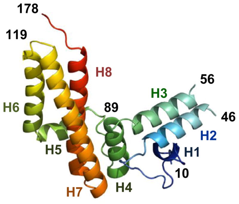
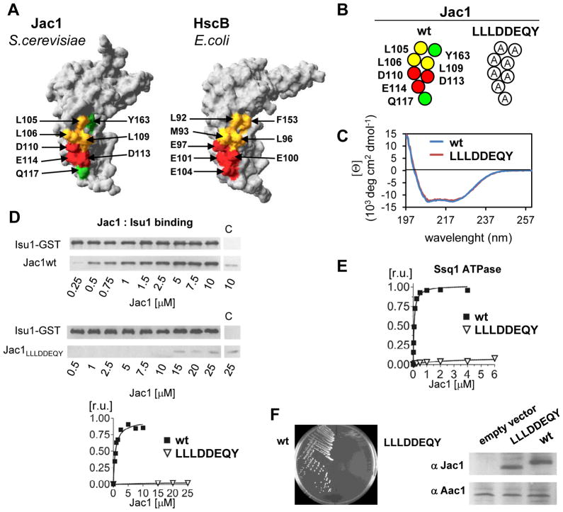
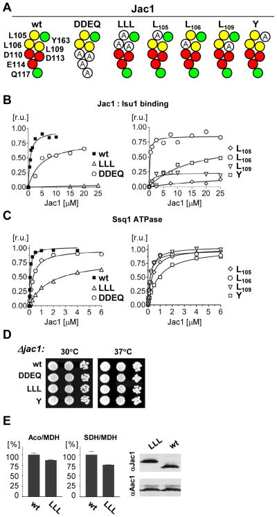
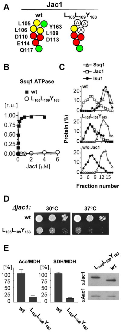
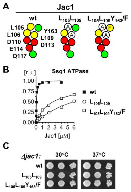
The ubiquitous mitochondrial J-protein Jac1, called HscB in Escherichia coli, and its partner Hsp70 play a critical role in the transfer of Fe-S clusters from the scaffold protein Isu to recipient proteins. Biochemical results from eukaryotic and prokaryotic systems indicate that formation of the Jac1-Isu complex is important for both targeting of the Isu for Hsp70 binding and stimulation of Hsp70's ATPase activity. However, in apparent contradiction, we previously reported that an 8-fold decrease in Jac1's affinity for Isu1 is well tolerated in vivo, raising the question as to whether the Jac1:Isu interaction actually plays an important biological role. Here, we report the determination of the structure of Jac1 from Saccharomyces cerevisiae. Taking advantage of this information and recently published data from the homologous bacterial system, we determined that a total of eight surface-exposed residues play a role in Isu binding, as assessed by a set of biochemical assays. A variant having alanines substituted for these eight residues was unable to support growth of a jac1-Δ strain. However, replacement of three residues caused partial loss of function, resulting in a significant decrease in the Jac1:Isu1 interaction, a slow growth phenotype, and a reduction in the activity of Fe-S cluster-containing enzymes. Thus, we conclude that the Jac1:Isu1 interaction plays an indispensable role in the essential process of mitochondrial Fe-S cluster biogenesis.


Figures

Fig. 1
Fig. 1. Saccharomyces cerevisiae Jac1 protein structure
Ribbon diagram of Jac1 protein. Rainbow coloring from blue to red indicates the N- to C-terminal positions of the residues in the model. The α-helices are numbered in corresponding colors. Numbers in black correspond to residue positions. The diagram was generated using Pymol (DeLano Scientific LL).
Fig. 2
Fig. 2. Alanine replacements of conserved charged and hydrophobic residues on the surface of the C-terminal domain of Jac1 result in defective Isu binding and inability to support cell growth
A. Corresponding residues are highlighted on the surface of protein crystal structures: (left) Jac1 (this report) and (right) HscB (PDB:1FPO) in the region implicated in interaction with Isu/IscU (see text). Yellow, hydrophobic residues; green, polar residues; red, negatively charged residues. B. A map of Jac1 surface residues and introduced substitutions to alanine in Jac1LLLDDEQY variant. C. CD spectra measured for purified wild-type (wt) Jac1 and Jac1LLLDDEQY as described in Materials and Methods. D. (top panel) Isu1-GST (2.5μM) and Jac1 wt or LLLDDEQY variant at the indicated concentrations were mixed to allow complex formation. Glutathione resin was added to pull-down the complex. Isu1-GST and Jac1 proteins were separated by SDS-PAGE and visualized by immunoblot analysis using antibodies specific for Isu1 and Jac1. As a control (c) Isu1-GST was omitted for 10 μM of Jac1 or Jac1LLLDDEQY as indicated. (bottom panel) Bound Jac1 was quantitated by densitometry. Values were plotted in Prism using 1:1 binding hyperbola to fit data for wt Jac1 (Kd = 0.69 ± 0.12 μM). Bmax was set to 1. E. Stimulation of Ssq1 ATPase activity by wt Jac1 or Jac1LLLDDEQY variant was measured in the presence of 0.5 μM Ssq1, 10 μM Isu1, 0.5 μM Mge1 and indicated concentrations of Jac1 proteins. Values were plotted in Prism using Michaelis-Menten hyperbolic equation to fit the data. The ATPase activity corresponding to maximal stimulation (MS) of wt Jac1 was set to 1. The concentration giving half-maximal stimulation was C05 = 0.065 ± 0.002 μM. F. (top) jac1-Δ cells harboring plasmid-borne copies of both wt JAC1(URA3 marked) and wt JAC1(HIS3 marked) or mutant jac1LLLDDEQY(TRP1 marked), as indicated, were plated on glucose-minimal medium containing 5-FOA, which selects for cells having lost the plasmid containing the wt copy of JAC1(URA3 marked). The plate was incubated at 30°C for 3 days. (bottom) Immunoblots of 0.1 optical density of cells lysates from indicated strains probed with antibodies specific to Jac1 and Aac1, a loading control. Empty vector- lysate of GAL-JAC1 strain, harbouring chromosomal copy of JAC1 under control of glucose repressible GAL-10 promoter, prepared following 64 hours of growth in glucose containing media. LLDDEQY- lysate of GAL-JAC1 strain transformed with plasmid harbouring copy of jac1LLDDEQY mutant under control of the native JAC1 promoter prepared after 64 hours of growth in glucose containing media. wt- lysate of wt yeast strain.
Fig. 3
Fig. 3. Importance of hydrophobic region for Isu binding in vitro
A. Maps of Jac1 surface residues and introduced substitutions to alanine in Jac1 variants. B. Isu1-GST (2.5μM) and Jac1 wt or Jac1 variants as indicated were mixed to allow complex formation. Glutathione resin was added to pull -down the complex and the samples were treated as described in Fig. 1. Bound Jac1 was quantitated by densitometry. Values were plotted in Prism using single binding hyperbola to fit data for wtJac1 (Bmax was set to 1; Kd =0.69 ± 0.12 μM), for Jac1DDEQ (Bmax = 0.81± 0.10 μM; Kd = 4.73 ±1.79 μM) and for Jac1L106(Bmax = 0.85±0.02 μM; Kd = 0.25±0.07 μM). C. Ssq1 ATPase stimulation was measured as described in Fig. 1. Values were plotted in Prism using Michaelis-Menten hyperbolic equation to fit the data. The ATPase activity corresponding to maximal stimulation (MS) of the wt Jac1 was set to 1. Kinetic parameters are listed in supplementary Table 3. D. jac1-Δ cells harboring plasmid-borne copies of wt JAC1 or mutant jac1, as indicated, were plated as 10-fold serial dilutions on glucose-rich medium and incubated at 30°C and 37°C for 3 days. E. (top) Aconitase activity (left) and succinate dehydrogenase activity (right) were measured in lysates of mitochondria isolated from jac1-Δ cells harboring plasmid -borne copies of wt JAC1or jac1LLL grown in glucose-minimal medium. As a standard, the non-FeS cluster-containing protein malate dehydrogenase (MDH) was measured. The ratio of activities of aconitase and MDH or SDH and MDH was calculated and expressed as percent of the ratio in wt mitochondrial extracts. Bars represents average values for three repeated measurements with presented error bars as S.D. (bottom) Protein concentrations of Jac1 and Aac1, a loading control, were determined in mitochondrial extracts from cells described in top panel. Mitochondrial extracts were separated by SDS-PAGE and proteins were visualized by immunoblot analysis using polyclonal antibodies as indicated.
Fig. 4
Fig. 4. Alanine replacement of L105, L109 and Y163 results in defective Fe-S cluster biogenesis in vivo
A. Map of Jac1 surface residues and introduced substitutions to alanine in Jac1L105L109Y163 B. Ssq1 ATPase stimulation was measured as described in Fig. 1. C. Isu1 binding to Ssq1 analyzed using glycerol gradient centrifugation. Purified proteins: Isu1 (2.5 μM), Jac1 (5 μM), Ssq1 (5 μM) were incubated, as indicated, in the presence of ATP (2 mM) in 70 μl reaction mixture prior to loading onto a gradient. Plots representing quantification of protein content obtained by densitometry after analysis of protein content of fractions by SDS-PAGE and silver staining. w/o Jac1- a negative control with no Jac1 in the reaction mixture. D. jac1-Δ cells harboring plasmid -borne copies of wt or mutant jac1, as indicated, were plated as 10-fold serial dilutions on glucose-rich medium and incubated at 30°C and 37°C for 3 days. E. (top) Aconitase (left) and succinate dehydrogenase (right) activities were measured in lysates of mitochondria isolated from jac1-Δ cells harboring plasmid -borne copies of wt JAC1or jac1L105L109Y163 grown in glucose-minimal medium. As a standard, the non-FeS cluster-containing protein malate dehydrogenase (MDH) was measured. The ratio of activities of aconitase and MDH or SDH and MDH was calculated and expressed as percent of the ratio in wt mitochondrial extracts. Bars represents average values for three repeated measurements with presented error bars as S.D. (bottom) Levels of Jac1 and Aac1, a loading control, in the mitochondrial extracts described in top panel were determined. Extracts were subjected to SDS-PAGE and proteins detected by immunoblot analysis using polyclonal antibodies as indicated.
Fig. 5
Fig. 5. Tyrosine 163 of Jac1 can be functionally replaced by Phenylalanine
A. Maps of Jac1 surface residues and introduced substitutions to alanine in Jac1 variants. B. jac1-Δ cells harboring plasmid-borne copies of wtJAC1 or jac1 mutant, as indicated, were plated as 10-fold serial dilutions on glucose-rich medium and incubated at 30°C and 37°Cfor 3 days. C. Ssq1 ATPase stimulation was measured as described in Fig. 1.

References

    1. Lill R, Muhlenhoff U. Maturation of iron-sulfur proteins in eukaryotes: mechanisms, connected processes, and diseases. Annu Rev Biochem. 2008;77:669–700. - PubMed
    1. Garland SA, Hoff K, Vickery LE, Culotta VC. Saccharomyces cerevisiae ISU1 and ISU2: members of a well-conserved gene family for iron-sulfur cluster assembly. J Mol Biol. 1999;294:897–907. - PubMed
    1. Gerber J, Muhlenhoff U, Lill R. An interaction between frataxin and Isu1/Nfs1 that is crucial for Fe/S cluster synthesis on Isu1. EMBO Rep. 2003;4:906–11. - PMC - PubMed
    1. Tsai CL, Barondeau DP. Human frataxin is an allosteric switch that activates the FeS cluster biosynthetic complex. Biochemistry. 2010;49:9132–9. - PubMed
    1. Craig EA, Marszalek J. A specialized mitochondrial molecular chaperone system: a role in formation of Fe/S centers. Cell Mol Life Sci. 2002;59:1658–65. - PMC - PubMed

Publication types

MeSH terms