. 2012 Feb 5;13(3):290-9.
doi: 10.1038/ni.2227.

The composition and signaling of the IL-35 receptor are unconventional

Affiliations

The composition and signaling of the IL-35 receptor are unconventional

Lauren W Collison et al. Nat Immunol. .

Abstract

Interleukin 35 (IL-35) belongs to the IL-12 family of heterodimeric cytokines but has a distinct functional profile. IL-35 suppresses T cell proliferation and converts naive T cells into IL-35-producing induced regulatory T cells (iTr35 cells). Here we found that IL-35 signaled through a unique heterodimer of receptor chains IL-12Rβ2 and gp130 or homodimers of each chain. Conventional T cells were sensitive to IL-35-mediated suppression in the absence of one receptor chain but not both receptor chains, whereas signaling through both chains was required for IL-35 expression and conversion into iTr35 cells. Signaling through the IL-35 receptor required the transcription factors STAT1 and STAT4, which formed a unique heterodimer that bound to distinct sites in the promoters of the genes encoding the IL-12 subunits p35 and Ebi3. This unconventional mode of signaling, distinct from that of other members of the IL-12 family, may broaden the spectrum and specificity of IL-35-mediated suppression.


Conflict of interest statement

COMPETING FINANCIAL INTERESTS

The authors declare competing financial interests. D.A.A.V., L.W.C. and K.M.V. have submitted patents that are pending and are entitled to a share in net income generated from licensing of these patent rights for commercial development. Furthermore, this work was supported in part by a sponsored research agreement with NovoNordisk to D.A.A.V.

Figures
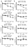
Figure 1
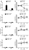
Figure 1. IL-12Rβ2 or gp130 are sufficient for IL-35-mediate suppression
(a–b) Tconv cells purified by flow sorting from wild-type (WT - C57BL/6), Il12rb2 −/−, CD4cre × Il6stfl/fl (abbreviated Il6stΔT), or Il12rb2 −/− × CD4cre × Il6stfl/fl (abbreviated IL-35RΔT) mice were activated with anti-CD3- + anti-CD28-coated beads for 3 days in the presence of indicated concentrations of IL-35-coated beads (isotype control or non-neutralizing anti-IL-35 mAb were incubated with IL-35 supernatant and then coupled with protein G beads) (a), or iTR35 in combination with neutralizing IL-35 mAb or isotype control mAb (b). Proliferation was determined by [3H]-thymidine incorporation. Counts per minute of Tconv cells activated alone, in the absence of any suppression, counts ranged from 22,000–65,000. Data represent the mean ± SEM of (a) 5 and (b) 3 (representative of 8) independent experiments. (* p < 0.05, ** p < 0.01 by unpaired t-test).
Figure 2
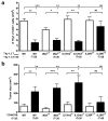
Figure 2. Both IL-12Rb2 and gp130 are required for Ebi3 and Il12a expression and iTr35 conversion
(a) Tconv cells were purified from WT, Il6st ΔT, Il12rb2 −/−, or IL-35R ΔT mice and activated in the presence of Tconv control or IL-35 supernatant, then assayed for Ebi3 and Il12a mRNA upregulation by qPCR. Results are normalized to Actb expression and scaled to WT cells stimulated with control supernatant. (b) Tconv purified by flow sorting from WT mice were activated with anti-CD3- + anti-CD28 coated beads for 3 days in the presence of iTrCon (cells differentiated in control supernatant) or iTr35 cells generated from WT, Il6st ΔT, Il12rb2 −/−, or IL-35RΔT mice. Proliferation was measured by [3H]-thymidine incorporation. CPM of Tconv cells activated alone ranged from 20,500–45,000. Data represent the mean of (a) 3 or (b) 3 [of 6] independent experiments (* p < 0.05, ** p < 0.01 by unpaired t-test).
Figure 3
Figure 3. IL-35R-deficient Tconv cells are resistant to IL-35-mediated suppression in vivo
(a) Homeostatic expansion was monitored by i.v. injection of Thy1.2+ Tconv cells from WT (C57BL/6), gp130 ΔT, Il12rb2 −/−, or IL-35RΔT mice alone or with Thy1.1+ iTr35 cells (as regulatory cells) into Rag1 −/− mice. Seven days after transfer, splenic T cell numbers were determined by flow cytometry. (b) Rag1 −/− mice received CD4+ and CD8+ T cells from WT (C57BL/6), gp130 ΔT, Il12rb2 −/−, or IL-35RΔT mice alone or with iTr35 cells via the tail vein on day −1 of the experiment. On day 0, all were injected with 120,000 B16 cells i.d. in the right flank. Tumor volume was measured 14 days as after inoculation (mm3). Data represent the mean ± SEM of (a) 5–12 and (b) 6–10 mice per group.
Figure 4
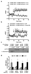
Figure 4. IL-12Rβ2 and gp130 associate in the presence of IL-35 to form three receptors
(a) Interaction of IL-12 with its receptor was determined using laser scanning spinning disc confocal microscopy combined with TIRF illumination and FRET-based detection. GFP-tagged IL-12Rβ1 and mCherry-tagged IL-12Rβ2 were transiently expressed on 293T cells. FRET excitation of the mCherry-IL-12Rβ2 acceptor following excitation of the GFP-IL-12Rβ1 donor was determined prior to and following the addition of IL-12 or IL-35 (arrow) by time lapsed imaging. FRET signals were mathematically corrected for donor bleed-through and direct excitation of the acceptor, and were normalized based on the FRET signal (nFRET) detected for the interval preceding addition of cytokine. (b) Similar analyses were performed using a GFP-tagged gp130 receptor as donor and mCherry-IL-12Rβ2 as acceptor following addition of IL-35 or control (arrow). Data represent the mean of (a) 2 and (b) 8 experiments, where significant increases in nFRET signals following addition of cytokine were considered to be significantly increased above control levels as noted: (a) * = p<0.001; (b) * = p<0.05. (c) Co-immunoprecipitation of receptor chains in response to cytokines. GFP- or mCherry-tagged receptor constructs were transiently co-expressed in 293T cells and then stimulated with IL-12 or IL-35. Cells were cross-linked with a cleavable cross-linker and lysed. Receptors were immunoprecipitated using anti-DsRed antibodies and interrogated for GFP signal by immunoblot analysis. (d) Densitometry scanning of four independent experiments as in b. GFP signal was normalized to the GFP signal from input lanes and scaled to vector-treated control (set to 1, * = p < 0.05, ** = p < 0.01, unpaired t-test).
Figure 5
Figure 5. IL35R chain expression can be induced IL-2 and IL-27
(a) Tconv cells were stimulated in the presence of varying doses of IL-2, IL-27, or neutralizing antibodies to IL-2 or IL-27p28. RNA was extracted and cDNA generated. Il6st, Il12rb1 and Il12rb2 mRNA expression was normalized to Actb expression and scaled to signal from naïve T cells (* = p < 0.05, ** = p < 0.01, *** = p < 0.001, unpaired t-test). (b,c) T cells were activated 48 h in the presence of indicated cytokines or anti-cytokine antibodies, rested 48 h, and restimulated with indicated concentrations of IL-35 for 3 days. Proliferation was determined by [3H]-thymidine incorporation. CPM of Tconv cells activated alone ranged from 31,000–75,000. Data represent the mean ± SEM of (a) 3, (b) 3, (c) 2 [of 6] independent experiments.
Figure 6
Figure 6. IL-35 signals through STAT1 and STAT4
(a) Tconv cells purified by flow sorting were treated with indicated cytokines for 30 min following 18h activation (αCD3-CD28) alone (IL-27) or activation (αCD3-CD28) in the presence of rIL-27 (IL-12 and IL-35). Intracellular staining of pSTATs was determined by flow cytometry (isotype control in grey). (b) Tconv cells purified by flow sorting were treated with IL-35 for indicated times following 24h activation (αCD3-CD28). Cells were lysed in cold RIPA buffer, resolved by SDS-PAGE and probed with anti-pSTAT Abs to identify STAT phosphorylation. (c) Tconv cells from wild-type (C57BL/6), gp130ΔT or Il12rb2−/− mice were stimulated with IL-35 and stained for pSTAT1 and pSTAT4 as in A (no stimulation in grey). (d) Immunoblot analysis of T cells blasts from WT (C57BL/6), gp130ΔT or Il12rb2−/− mice stimulated with IL-35 for 30 min and probed as in b. (e) Tconv cells were purified by flow sorting from WT (C57BL/6 or Balb/c), Stat1−/− (C57BL/6 background), Stat3−/− (C57BL/6 background), or Stat4−/− (Balb/c background) mice. Tconv cells were activated with anti-CD3- + anti-CD28-coated latex beads for 3 days in the presence of indicated concentrations of IL-35. Proliferation was determined by [3H]-thymidine incorporation. Counts per minute of Tconv cells activated alone ranged from 24,000–91,000. Data represent the mean ± SEM of (a,b,e) 3–8, (c) 4, and (d) 3 independent experiments.
Figure 7
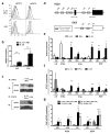
Figure 7. IL-35 utilizes a STAT1:STAT4 heterodimer to mediate Ebi3 and Il12a promoter interaction
(a) Tconv cells were activated 48 h in the presence of IL-2 and IL-27, rested for 5 d in IL-2, serum starved for 3 h on ice, and stimulated with control supernatant, IL-12 + IFN-γ, or IL-35. Cells were stained intracellularly for pSTAT1 and pSTAT4. Shaded histograms represents control supernatant treatment. (b) Tconv cells activated for 48 h in the presence of control supernatant, IL-35, or IL-12 + IFN-γ were interrogated for Ebi3 and Il12a mRNA by qPCR. Values represent fold increase over control supernatant normalized to Actb expression (* = p < 0.05, ** = p < 0.01, unpaired t-test). (c) Tconv cells were stimulated as in a and lysed. Lysates were precleared, then STAT1 or STAT4 was immunoprecipitated. Eluates were immunoblotted for STAT1 and STAT4. (d) Schematic diagram of the mouse Il12a and Ebi3 promoters. Eight STAT binding sites were identified; six are interrogated here. (e) ChIP analysis of STAT4 binding sites in the Il12a and Ebi3 promoters. Tconv cells were activated with anti-CD3, anti-CD28, and IL-2 for 48 h, expanded in IL-2 for 48 h, serum starved 3 h, then stimulated with control supernatant, -IL-12, IFN-γ, or IL-35 for 90 min. Chromatin was prepared from 15 × 106 T cells per stimulation and subjected to STAT4 ChIP. ChIP DNA was assayed using real-time PCR. Primers to Irf1 and Il18ra promoter sites were used as controls. Results represent fold increase over control protein treated cells, which are scaled using isotype-IgG ChIP and input (* = p < 0.05, ** = p < 0.01, unpaired t-test IL-12 vs. IL-35). (f) ChIP analysis as in (e), but for STAT1 (* = p < 0.05, ** = p < 0.01, unpaired t-test, IFN-γ vs IL-35). (g) Expanded Tconv cells (30 × 106) were prepared as in e. The eluted chromatin from STAT4 or STAT1 ChIP was then subjected to a second IP (reChIP). Analyses were restricted to loci in which both STAT1 and STAT4 ChIP signals were enriched following IL-35 stimulation in e and f (* = p < 0.05, ** = p < 0.01, unpaired t-test, IL-12+IFN-γ vs. IL-35). Data are representative (a,c) or are the mean of 3 (b,f), 5 (e) independent experiments, or 2 [of 3] (g) independent experiments.
Figure 8
Figure 8. Structural models of the IL-12 family of cytokine receptor complexes
(a) Structure of the complete ectodomain receptor signaling complex for IL-6 with IL-6Ra and gp130 derived from electron microscopic and crystallographic data, , . Sites 1, 2 and 3 are indicated on the model. (b) Models of potential IL-35 signaling complexes based on the IL-6 complex demonstrating alternative arrangements of the receptors as signaling-competent homo- or hetero-meric dimers. (c) Models of ectodomain complexes of IL-27, IL-12, and IL-23 based on the IL-6 complex. Models were constructed, visualized and rendered in Pymol (Schrödinger LLC The PyMOL Molecular Graphics System, Version 1.2r3pre).

References

    1. Vignali DA, Collison LW, Workman CJ. How regulatory T cells work. Nat Rev Immunol. 2008;8:523–532. - PMC - PubMed
    1. Zou W. Regulatory T cells, tumour immunity and immunotherapy. Nat Rev Immunol. 2006;6:295–307. - PubMed
    1. O’Shea JJ, Paul WE. Regulation of T(H)1 differentiation--controlling the controllers. Nat Immunol. 2002;3:506–508. - PubMed
    1. Artis D, et al. The IL-27 receptor (WSX-1) is an inhibitor of innate and adaptive elements of type 2 immunity. J Immunol. 2004;173:5626–5634. - PubMed
    1. Awasthi A, et al. A dominant function for interleukin 27 in generating interleukin 10-producing anti-inflammatory T cells. Nat Immunol. 2007;8:1380–1389. - PubMed

Publication types

MeSH terms