. 2012 Feb 5;13(3):237-45.
doi: 10.1038/ni.2225.

A CD74-dependent MHC class I endolysosomal cross-presentation pathway

Affiliations

A CD74-dependent MHC class I endolysosomal cross-presentation pathway

Genc Basha et al. Nat Immunol. .

Abstract

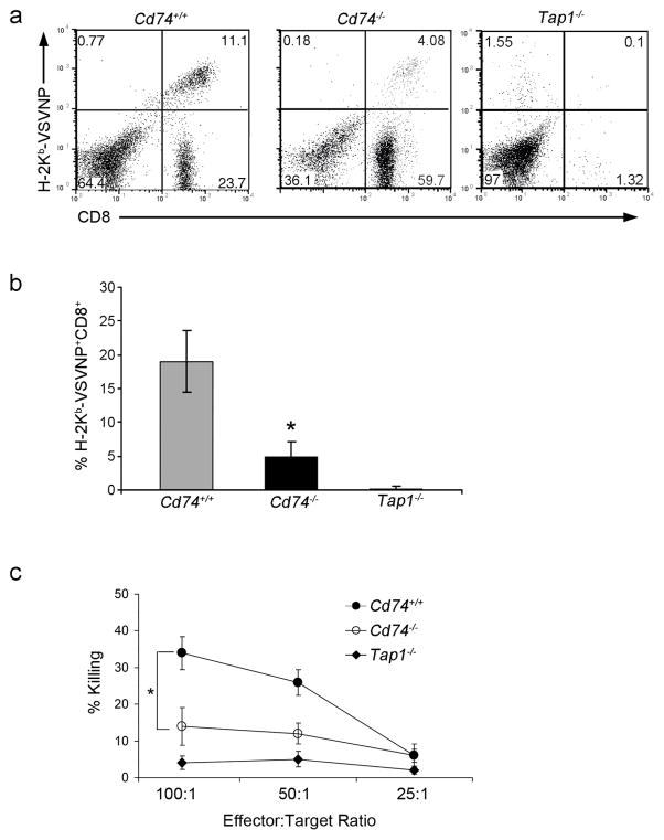
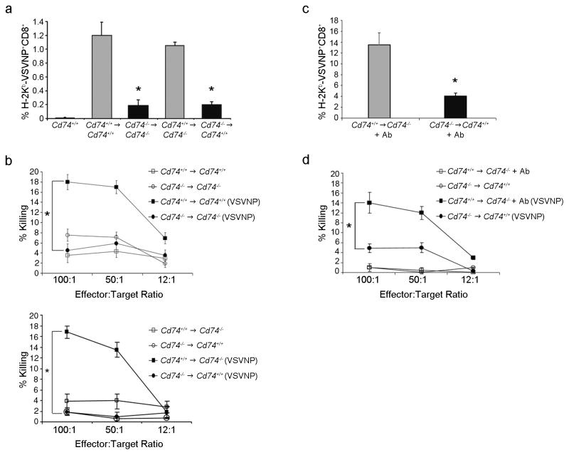
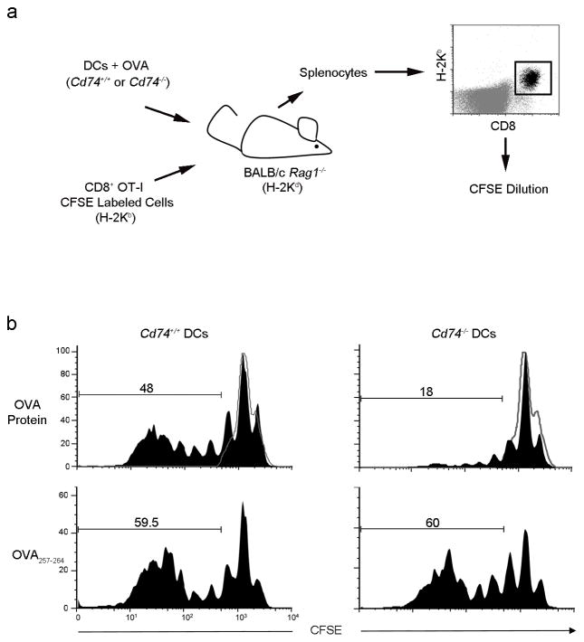
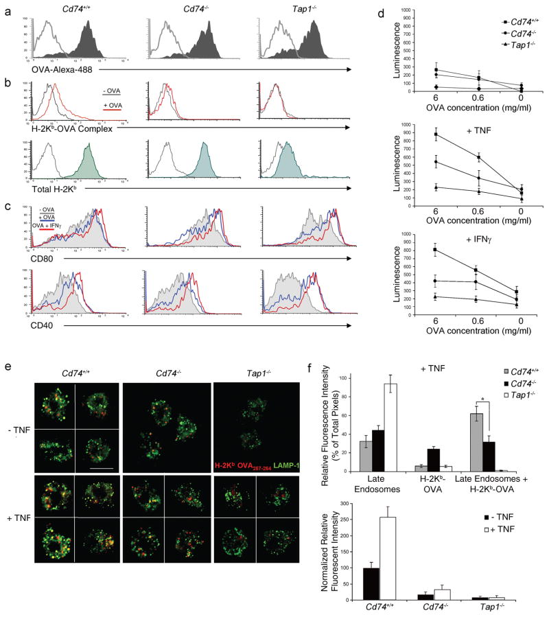
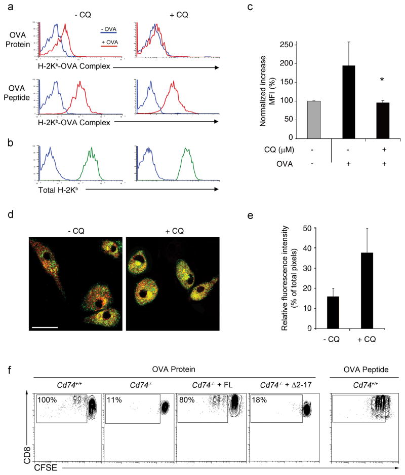
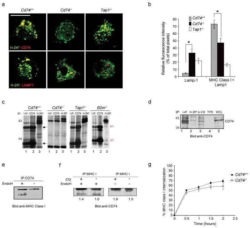
Immune responses are initiated and primed by dendritic cells (DCs) that cross-present exogenous antigen. The chaperone CD74 (invariant chain) is thought to promote DC priming exclusively in the context of major histocompatibility complex (MHC) class II. However, we demonstrate here a CD74-dependent MHC class I cross-presentation pathway in DCs that had a major role in the generation of MHC class I-restricted, cytolytic T lymphocyte (CTL) responses to viral protein- and cell-associated antigens. CD74 associated with MHC class I in the endoplasmic reticulum of DCs and mediated the trafficking of MHC class I to endolysosomal compartments for loading with exogenous peptides. We conclude that CD74 has a previously undiscovered physiological function in endolysosomal DC cross-presentation for priming MHC class I-mediated CTL responses.


Figures

Figure 1
Figure 1. Cd74−/− mice generate weak antiviral primary immune responses
Cd74+/+, Cd74−/− and Tap1−/− mice were infected with a low titer of VSV (2 × 10⁵ TCID50 or dose that infects 50% of a tissue culture cell monolayer/mouse). (a) Six days following viral infection, splenocytes were isolated and after a 5-day stimulation with VSVNP52-59, the number of VSVNP52-59-specific CD8+ T cells generated was assessed. Percentages of VSVNP52-59-specific CD8+ T cells in representative mice are shown. (b) Mean percentages (± SD) of H-2Kb-VSVNP52-59-specific CD8+ T cells of three mice are shown. (c) Standard 51Cr-release assays were performed using CTLs generated following VSV infection and in vitro boosting. Error bars represent SD. Data are representative of at least three separate experiments. * p<0.05.
Figure 2
Figure 2. The deficiency of Cd74−/− mice to elicit primary immune responses resides in their APCs and is independent of CD4+ T cells
(a) Chimeras were injected with 1 × 10⁵ TCID50 VSV and splenocytes were assessed for the generation of VSVNP52-59 -specific CD8+ cells. The mean percentage (± SD) of three mice assayed following in vitro boosting with VSV NP52-59 peptide is shown. (b) Cytotoxicity assays were performed using CTLs generated following in vitro boosting. Data are representative of at least three separate experiments. (c) Cd74+/+Cd74−/− chimeras were depleted of CD4+ cells by intravenous injection of a CD4 (GK1.5) antibody (+Ab) then assessed for immune function. Mice chimeras infected with VSV were evaluated for the generation of H-2Kb-VSVNP52-59-specific CD8+ T cells. The mean percentage (± SD) of tetramer+CD8+ cells in the spleen of three mice is shown. (d) The lytic activity of the immune response was also assessed. Error bars represent SD. * p < 0.05.
Figure 3
Figure 3. Cd74−/− DCs are unable to cross-present cell-associated antigens in vivo to prime antigen-specific CD8+ T cells
(a) OVA protein or OVA257-264 pulsed Cd74−/− or Cd74+/+ BMDCs were injected with purified CD8+ OT-I CFSE-labeled T cells into Rag1−/− mice on a BALB/c background. Three days later, H-2KbCD8+ T cells were assessed for proliferation. (b) Black histograms represents proliferating OT-I derived T cells from the spleens of representative mice (n=3). Grey histograms represent unproliferating OT-I T cells.
Figure 4
Figure 4. Cross-presentation and cross-priming is defective in Cd74−/−-derived DCs
(a) BMDCs were incubated with OVA-Alexa-488 and uptake (shaded) was assessed over background (grey) by flow cytometry. (b) Formation of H-2Kb-OVA257-264 complexes on splenic DCs with (red) or without (grey) incubation with soluble OVA as well as total H-2Kb (shaded) above background (grey) was measured by flow cytometry. (c) Costimulatory receptors CD80 and CD40 on BMDCs were assessed by flow cytometry following incubation with OVA (blue), OVA+IFNγ (red) or media alone (grey). (d) Spleen-derived DCs were incubated with soluble OVA as indicated, in the presence of GM-CSF or GM-CSF and TNF or IFN-γ. Activation of B3Z T cells was measured using a chemiluminescent assay. Data depict means (± SD) of triplicate samples for each OVA concentration. Similar results were observed in 3 separate experiments. (e) Mature spleen-derived DCs incubated with OVA were costained with H-2Kb-OVA257-264 specific antibody (red) and LAMP-1 (green). The figure shows optically merged images representative of the majority of cells examined by ICM. Scale bar, 5 μm. (f) Quantitative assessment was performed comparing H-2Kb-OVA257-264 colocalization with LAMP-1+ late endosomes with or without TNF (top panel). Quantitative assessment comparing fluorescence between Cd74+/+, Cd74−/− and Tap1−/− DCs with TNF treatment (bottom panel). For each analysis >20 DCs per mouse strain were examined. Graph depicts normalized individual color pixel percentages per total pixels ± SD. * p < 0.05.
Figure 5
Figure 5. Inhibition of CD74-mediated MHC I trafficking in DCs by treatment with chloroquine
(a) BMDCs were treated with CQ and the formation of H-2Kb-OVA257-264 complexes on DCs following incubation with soluble OVA (red; top panel) or OVA peptide (red; bottom panel) compared to media alone (blue) was measured by flow cytometry. (b) Total H-2Kb (green) above background (blue) was also assessed. (c) The fold increase in surface H-2Kb-OVA257-264 complexes following incubation with soluble OVA was quantified. Graphs depict normalized increases in mean fluorescence intensity (MFI) ± SD. * p < 0.05. (d) Mature BMDCs treated with CQ were costained with H-2Kb (red) and CD74 (green) antibody. The figure shows optically merged images representative of the majority of cells examined by ICM. Scale bar, 5 μm. (e) Quantitative assessment comparing H-2Kb colocalization with CD74. Graph depicts normalized individual color pixel percentages divided per total pixels ± SD. (f) Cd74−/− BMDCs reconstituted with full length (FL) CD74 or truncated (Δ2–17) CD74 lacking the endolysosomal trafficking motif were incubated with soluble OVA and assessed for the ability to induce proliferation in CFSE-labeled OT-I cells. Percentages represent the proportion of proliferating OT-I cells normalized to Cd74+/+ controls.
Figure 6
Figure 6. CD74 controls MHC class I ER-to-endolysosome trafficking in DCs
(a) Mature splenic DCs were stained with H-2Kb (green) and CD74 (red) or LAMP1 (red) antibodies. Representative images as examined by ICM are shown. Scale bar, 5 μm. (b) Quantitative assessment of MHC class I in LAMP+ compartments was performed (50 DCs per mouse strain). Graphs depicts individual color pixel percentages per total pixels (mean ± SD) (c). Immunoprecipitation using H-2Kb, I-A/I-E and CD74 antibodies was performed on [35S]methionine-labeled BMDC. The CD74 41 and 31 k-Da protein bands are indicated. (d) Immunoprecipitation with antibodies against I-Ab, H-2Kb (conformationally dependent), H-2Kb cytoplasmic domain (e-VIII; conformationally independent) or transferrin receptor (TFR) was performed with Cd74+/+ DC lysates. The identity of the co-immunoprecipitated proteins was confirmed by blotting with a CD74 antibody. Whole cell lysate (WCL) was blotted as a control. (e) DC cell lysates were immunoprecipitated with a CD74 antibody and digested with endoglycosidase H. Immunoblotting with a MHC class I antibody was used to assess the MHC class I fraction precipitated by CD74 antibody and to visualize the acquisition of EndoH resistance of the MHC class I subset interacting with CD74. (f) Cells were treated with CQ to enhance CD74 and MHC I interaction and cell lysates were immunoprecipitated with a MHC class I antibody. Western blotting with a CD74 antibody was used to assess the CD74 protein fraction associating with MHC class I. (g) DCs labeled with a H-2Kb antibody were evaluated overtime for MHC class I internalization measured by flow cytometry as a reduction in mean fluorescence intensities over time. * p < 0.05.

Comment in

References

    1. Guagliardi LE, et al. Co-localization of molecules involved in antigen processing and presentation in an early endocytic compartment. Nature. 1990;343:133–139. - PubMed
    1. Kovacsovics-Bankowski M, Rock KL. A phagosome-to-cytosol pathway for exogenous antigens presented on MHC class I molecules. Science. 1995;267:243–246. - PubMed
    1. Ackerman AL, Kyritsis C, Tampe R, Cresswell P. Early phagosomes in dendritic cells form a cellular compartment sufficient for cross presentation of exogenous antigens. Proc Natl Acad Sci U S A. 2003;100:12889–12894. - PMC - PubMed
    1. Guermonprez P, et al. ER-phagosome fusion defines an MHC class I cross-presentation compartment in dendritic cells. Nature. 2003;425:397–402. - PubMed
    1. Houde M, et al. Phagosomes are competent organelles for antigen cross-presentation. Nature. 2003;425:402–406. - PubMed

Publication types

MeSH terms

Substances