. 2012 Feb 5;18(3):396-404.
doi: 10.1038/nm.2629.

Activin-like kinase 3 is important for kidney regeneration and reversal of fibrosis

Affiliations

Activin-like kinase 3 is important for kidney regeneration and reversal of fibrosis

Hikaru Sugimoto et al. Nat Med. .

Abstract

Molecules associated with the transforming growth factor β (TGF-β) superfamily, such as bone morphogenic proteins (BMPs) and TGF-β, are key regulators of inflammation, apoptosis and cellular transitions. Here we show that the BMP receptor activin-like kinase 3 (Alk3) is elevated early in diseased kidneys after injury. We also found that its deletion in the tubular epithelium leads to enhanced TGF-β1-Smad family member 3 (Smad3) signaling, epithelial damage and fibrosis, suggesting a protective role for Alk3-mediated signaling in the kidney. A structure-function analysis of the BMP-Alk3-BMP receptor, type 2 (BMPR2) ligand-receptor complex, along with synthetic organic chemistry, led us to construct a library of small peptide agonists of BMP signaling that function through the Alk3 receptor. One such peptide agonist, THR-123, suppressed inflammation, apoptosis and the epithelial-to-mesenchymal transition program and reversed established fibrosis in five mouse models of acute and chronic renal injury. THR-123 acts specifically through Alk3 signaling, as mice with a targeted deletion for Alk3 in their tubular epithelium did not respond to therapy with THR-123. Combining THR-123 and the angiotensin-converting enzyme inhibitor captopril had an additive therapeutic benefit in controlling renal fibrosis. Our studies show that BMP signaling agonists constitute a new line of therapeutic agents with potential utility in the clinic to induce regeneration, repair and reverse established fibrosis.


Figures
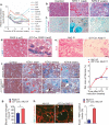
Figure 1
Figure 1. Alk3 and BMP7 expression inversely correlate in kidneys developing progressive injury and fibrosis
(a) Gene expression of indicated genes in kidneys of mice before (day 0) and after nephrotoxic serum nephritis (NTN) induction (1 week, 3 weeks, 6 weeks and 9 weeks after induction). (b) Representative MTS staining of control kidneys and kidneys from mice at one and six weeks following NTN. Scale bar: 50 μm. (c) Representative picture of kidneys from control mice and from mice at one and six weeks following NTN labeled with antibodies against phosphorylated Smad1 (p–Smad1). Scale bar: 50 μm. (d) Beta–galactosidase substrate staining of kidneys of control mice (left) and γGT–Cre; R26R–Rosa–LSL–LacZ reporter mice (right) and eosin counter stain. Scale bar: 50 μm, arrows: positive LacZ staining. (e) Representative picture of kidneys from control Alk3 f/f (left) and γGT–Cre; Alk3 f/f mice (right) labeled with antibodies against Alk3. Scale bar: 100 μm. (f) Representative MTS staining (left) of NTN kidney from γGT–Cre; Alk3 f/f mice (n = 6) and littermate control mice (Alk3 f/f, n = 4) and morphometric quantification (right) of fibrosis in NTN kidney from control (Alk3 f/f) and γGT–Cre; Alk3 f/f mice. Scale bar: 50 μm. (g) Blood urea nitrogen measurement 60 days following NTN in γGT–Cre; Alk3 f/f mice (n = 5) and littermate control mice (n = 3). (h) Representative images for E–cadherin / FSP1 immunolabeling of kidneys from control (Alk3 f/f) and γGT–Cre; Alk3 f/f mice (left) and percent of E–cadherin / FSP1 double positive tubule (right). Scale bar: 25 μm. Data are expressed as mean ± s.e.m. * : P < 0.01.
Figure 2
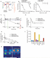
Figure 2. Synthesis and pharmacokinetics of THR–123
(a) Structure of BMP7 and THR–123. (b) Radio–ligand receptor competitive binding assays of THR–123 for Alk3 (left) and Alk6 (right) extra–cellular domain (ECD) (expressed as a fusion protein with Fc domain). (c) Stability of THR–123 in plasma and whole blood of rat following i.v. injection of 125I–THR–123, data are expressed as the ratio of THR–123 concentration at the time of harvest (conc. at t) over THR–123 concentration at baseline (conc. at t = 0). (d–e) alpha phase (d) and beta phase (e) of THR–123 clearance in systemic circulation of rats following i.v. injection of 125I–THR–123. (f). Tissue distribution of 125I–THR–123 six hours after i.v. administration. (g) Visualization of radioactivity of orally administrated 125I–THR–123 in rat kidneys at 1, 3, 6 and 24 hours after administration. Scale bar: 15 mm. Data are expressed as mean ± s.e.m.
Figure 3
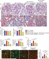
Figure 3. THR–123 reverses renal fibrosis in mice with nephrotoxic serum nephritis
(a) Representative MTS staining of kidney from untreated control mice (n = 5) (left panel); six weeks following NTN (n = 6, NTN, 6 weeks) (middle left panel); nine weeks post NTN (NTN, 9 weeks) (middle right panel); and nine weeks NTN with THR–123 administered starting at week 6 (NTN, 9 weeks + THR–123 (6–9 weeks)) (left panel), Scale bar: 40 μm. (b) Morphometric analysis from control mice (n = 5), mice after 1 (n = 6), 3 (n = 8), and six weeks following induction of NTN (n = 6), and mice at nine weeks following NTN with THR–123 administered starting six weeks following NTN (THR–123 (6–9 weeks) (n = 6)), assessing percent of glomerulosclerosis score (left), tubular atrophy index (middle), and fibrosis index (left). (c) Blood urea nitrogen measurement for mice six weeks following NTN (n = 3), nine weeks (n = 5), and nine weeks following NTN with THR–123 administered starting at week 6 following NTN (n = 5). (d) Representative images of E–cadherin / FSP1 immunolabeling of kidney from control untreated mice (left panel), six weeks following NTN (middle left panel), nine weeks (middle right panel), and nine weeks following NTN with THR–123 administered starting at week 6 following NTN (right panel) and percent of E–cadherin / FSP1 double positive tubule assessed by counting the number of double–labeled tubules (right). Scale bar: 25 μm. Data are expressed as mean ± s.e.m. ** : P < 0.01, * : P < 0.05.
Figure 4
Figure 4. THR–123 reverses fibrosis associated with diabetic nephropathy
(a) Representative histological PAS (upper panels) and MTS (lower panels) staining of kidney sections of control mice (n = 5, left panel), five months following streptozotocin–induced diabetic nephropathy (DN) (n = 6, 2nd panel from the left), six months following DN (n = 10, middle panel), six months following DN and treated with BMP7 from one to six months following DN (n = 4, 4th panel) or THR–123 from five to six month following DN (n = 9, right panel). Scale bar upper panels: 10 μm, scale bar lower panels: 50 μm. (b) Morphometric analysis of glomerular surface area (top left), mesangial matrix (top right), tubular atrophy (bottom left) and relative interstitial volume (bottom right). (c) Blood urea nitrogen measurement of control mice, five months following DN, six months following DN, six months following DN and treated with BMP7 from one to six months and six months following DN and treated with THR–123 from one to six months, and urine albumin over urine creatinine ratio of indicated experimental groups. (d) Representative images of E–cadherin / FSP1 immunolabeling of kidney of indicated experimental groups and percent of E–cadherin / FSP1 double positive tubule assessed by counting the number of double–labeled tubules. Scale bar 25 μm. Data are expressed as mean ± s.e.m. * : P < 0.01.
Figure 5
Figure 5. A combination of captopril and THR–123 inhibits progression of fibrosis associated with advanced diabetic nephropathy
(a) Representative histological PAS (upper panels) and MTS (lower panels) staining of kidney sections of mice seven months following streptozotocin–induced diabetic nephropathy (DN) (n = 2), eight months following DN induction (n = 3), eight months following DN induction and treated with captopril (CPR, n = 3), and eight months following DN induction and treated with combination of CPR and THR–123 (n = 4). Scale bar upper panels: 10 μm, scale bar lower panels: 50 μm. (b) Morphometric analysis of glomerular surface area (top left), mesangial matrix (top right), tubular atrophy (bottom left) and relative interstitial volume (bottom right) of indicated experimental groups. (c) Blood urea nitrogen measurement in indicated experimental groups. (d) Representative images of E–cadherin / FSP1 immunolabeling of kidney from indicated experimental groups and percent of E–cadherin / FSP1 double positive tubules. Scale bar: 25 μm. Data are expressed as mean ± s.e.m. ** : P < 0.01, * : P < 0.05, # : P = 0.08, ns: not significant.
Figure 6
Figure 6. Alk3 is a functional receptor for the activity of THR–123
(a) Representative MTS staining picture of Alk3 f/f and γGTCre; Alk3 f/f mice six weeks after NTN and treated with PBS or THR–123 (n = 4 in each group). And morphometric analyses of interstitial fibrosis in kidneys of mice the indicated experimental groups. Scale bar: 50 μm. (b) Representative images of F4/80 immunofluorescence in the indicated experimental groups and quantitation of the number of F4/80+ macrophages (Mϕ) per field of view. Scale bar: 25 μm. (c) Representative images of E–cadherin / FSP1 immunolabeling of kidney from indicated experimental groups and percent of E– cadherin / FSP1 double positive tubules. Scale bar: 25 μm. (d) Blood urea nitrogen measurement in six and nine weeks of NTN in indicated experimental groups (all n = 4). Data are expressed as mean ± s.e.m. ** : P < 0.01, * : P < 0.05. ns: not significant.

Comment in

References

    1. Zeisberg M, Kalluri R. The role of epithelial-to-mesenchymal transition in renal fibrosis. J Mol Med. 2004;82:175–181. - PubMed
    1. Zeisberg M, et al. BMP-7 counteracts TGF-beta1-induced epithelial-to mesenchymal transition and reverses chronic renal injury. Nat Med. 2003;9:964–968. - PubMed
    1. Zeisberg EM, et al. Endothelial-to-mesenchymal transition contributes to cardiac fibrosis. Nat Med. 2007;13:952–961. - PubMed
    1. Miyazono K, Maeda S, Imamura T. BMP receptor signaling: transcriptional targets, regulation of signals, and signaling cross-talk. Cytokine Growth Factor Rev. 2005;16:251–263. - PubMed
    1. Simic P, Vukicevic S. Bone morphogenetic proteins: from developmental signals to tissue regeneration. Conference on bone morphogenetic proteins. EMBO Rep. 2007;8:327–331. - PMC - PubMed

Publication types

MeSH terms

Substances