. 2012 Sep;61(4):566-74.
doi: 10.1016/j.neuint.2012.01.013. Epub 2012 Jan 28.

The glutamate transporter, GLAST, participates in a macromolecular complex that supports glutamate metabolism

Affiliations

The glutamate transporter, GLAST, participates in a macromolecular complex that supports glutamate metabolism

Deborah E Bauer et al. Neurochem Int. 2012 Sep.

Abstract

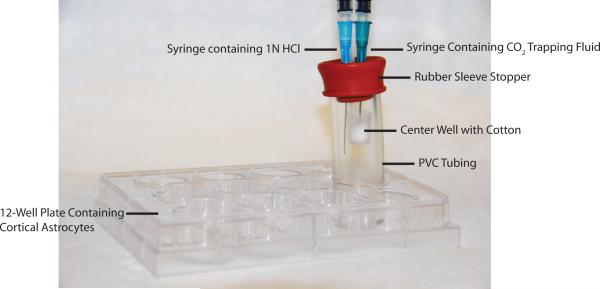
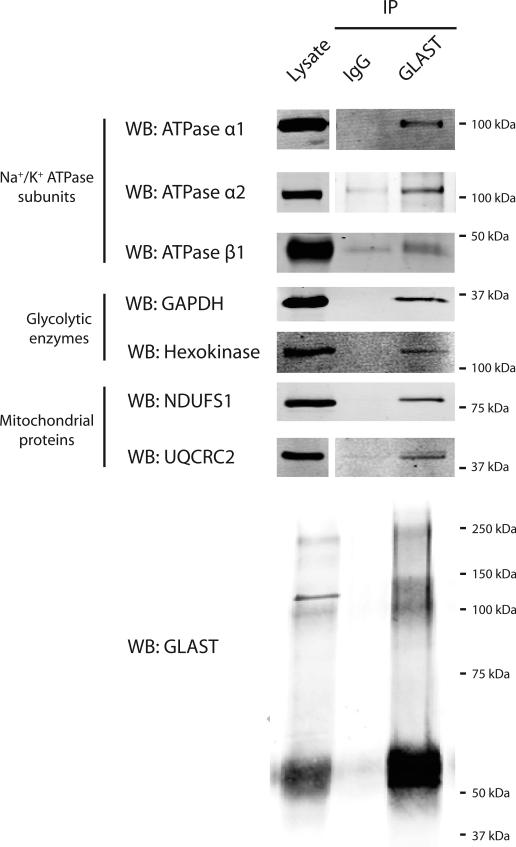
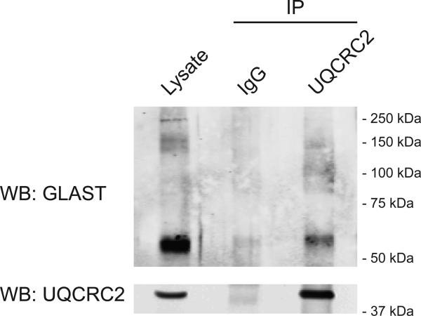
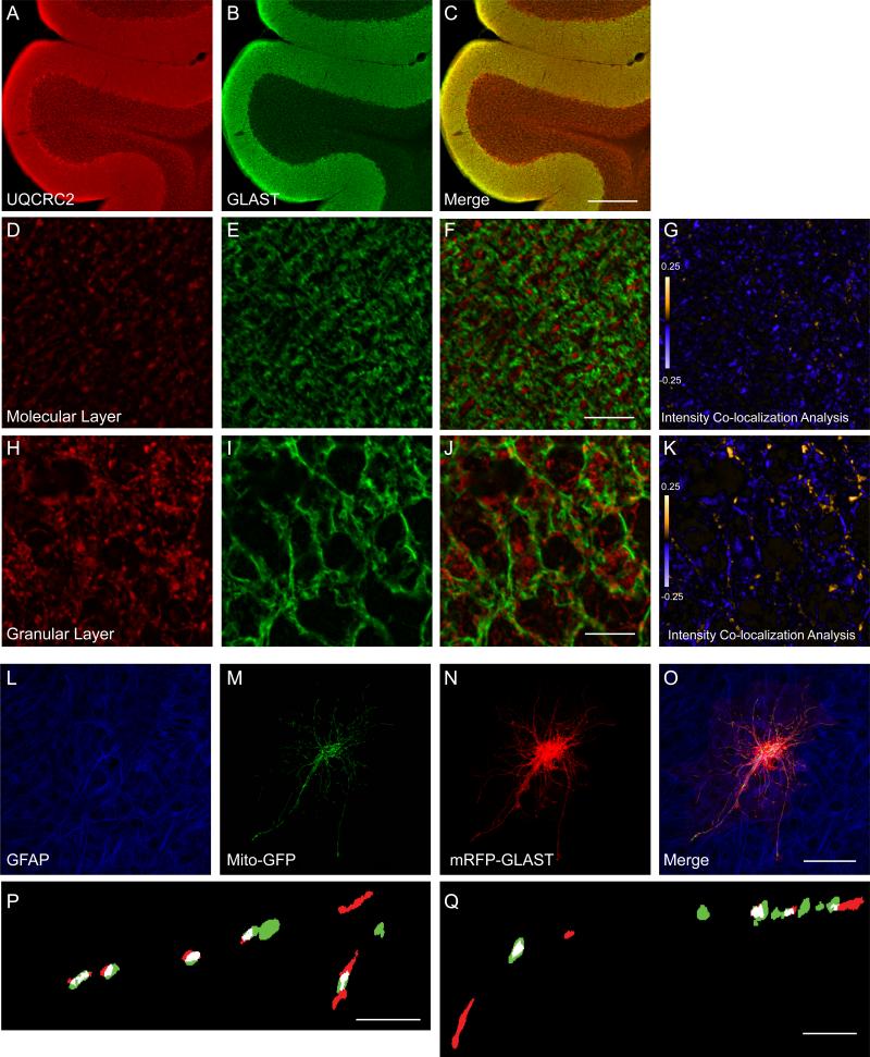
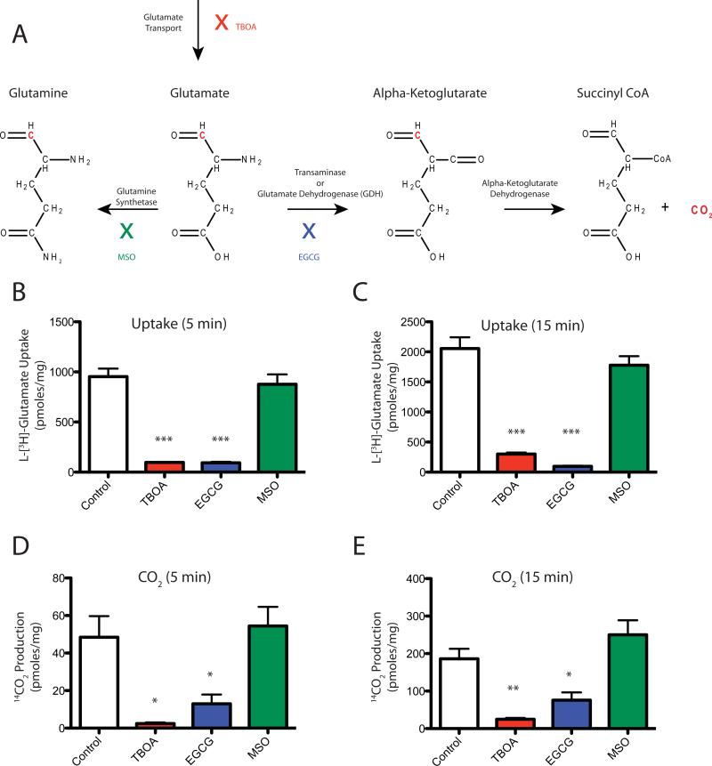
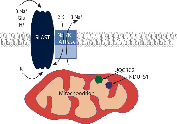
GLAST is the predominant glutamate transporter in the cerebellum and contributes substantially to glutamate transport in forebrain. This astroglial glutamate transporter quickly binds and clears synaptically released glutamate and is principally responsible for ensuring that synaptic glutamate concentrations remain low. This process is associated with a significant energetic cost. Compartmentalization of GLAST with mitochondria and proteins involved in energy metabolism could provide energetic support for glutamate transport. Therefore, we performed immunoprecipitation and co-localization experiments to determine if GLAST might co-compartmentalize with proteins involved in energy metabolism. GLAST was immunoprecipitated from rat cerebellum and subunits of the Na(+)/K(+) ATPase, glycolytic enzymes, and mitochondrial proteins were detected. GLAST co-localized with mitochondria in cerebellar tissue. GLAST also co-localized with mitochondria in fine processes of astrocytes in organotypic hippocampal slice cultures. From these data, we hypothesized that mitochondria participate in a macromolecular complex with GLAST to support oxidative metabolism of transported glutamate. To determine the functional metabolic role of this complex, we measured CO(2) production from radiolabeled glutamate in cultured astrocytes and compared it to overall glutamate uptake. Within 15 min, 9% of transported glutamate was converted to CO(2). This CO(2) production was blocked by inhibitors of glutamate transport and glutamate dehydrogenase, but not by an inhibitor of glutamine synthetase. Our data support a model in which GLAST exists in a macromolecular complex that allows transported glutamate to be metabolized in mitochondria to support energy production.


Figures

Figure 1
Figure 1. Device used to capture CO2
Astrocytes were cultured in 12-well plates. ACSF containing L-[1-14C]-glutamate was added to the well, and the well was immediately plugged with PVC tubing and a stopper. A center well containing cotton was suspended by the stopper. The reaction was stopped by injection of HCl through a 1.5 inch needle, and then CO2 was trapped by injection of a scintillation fluid-compatible base into the center well with a 1 inch needle.
Figure 2
Figure 2. Immunoprecipitation of GLAST from cerebellar lysates
Anti-GLAST antibody or IgG was used for immunoprecipitations from rat cerebellar lysates (750 μg protein). In all cases, the lysates were analyzed in the same Western blots. Some blots were cropped. Immunoprecipitation of GLAST was confirmed in every immunoprecipitation and one example is presented. Data are representative of at least 3 independent experiments.
Figure 3
Figure 3. Immunoprecipitation of UQCRC2 from cerebellar lysates
Anti-UQCRC2 antibody or IgG was used for immunoprecipitations from rat cerebellar lysates (500 μg protein). Lysates were analyzed in the same Western blots. Data are representative of 2 independent experiments. Note: GLAST monomers migrate at about 60 kDa.
Figure 4
Figure 4. Co-localization of GLAST with mitochondria in cerebellum (A-K) and in biolistically transduced astrocytes in organotypic hippocampal slice cultures (L- Q)
A-C. Representative images from adult rat cerebellum immunostained with antibodies against UQCRC2 (A; red) and GLAST (B; green). C. Merged image of GLAST and UQCRC2 immunofluorescence. Scale bar = 300 μm. D-K. Higher magnification views of the molecular layer (D, E) and granular layer (H, I) of the cerebellum immunostained for UQCRC2 (red; D, H) and GLAST (green; E, I) and merged (F, J). Scale bar = 10 μm. G, K. Pseudocolor representatations of PDM values for the image pairs (D:E and H:I), where pixel intensity is equal to the PDM value at that location. Images with positive PDM value are displayed in yellow, while those with negative values are displayed in violet. L-Q. Co-localization of mitochondria and GLAST in the processes of astrocytes in slice culture. Representative images of astrocytes in slice culture immunostained for glial fibrillary acidic protein (L; GFAP; blue) and transfected with the cDNAs encoding Mito-GFP (M, green) and mRFP-GLAST (N, red) and a merged image (O). P, Q. Binary, higher magnification view of astrocyte processes. Overlapping pixels are indicated in white.
Figure 5
Figure 5. Stoichiometry of CO2 production from L-[1-14C] glutamate in cortical astrocyte cultures
Glutamate uptake and CO2 production were measured in cortical astrocyte cultures at 5 or 15 minutes in the absence or presence of TBOA (1 mM), EGCG (1 mM), or MSO (5 mM). A. Potential Glutamate Pathways: Glutamate that has been transported into astrocytes can be converted into glutamine by glutamine synthetase or converted to alpha-ketoglutarate by transaminase or glutamate dehydrogenase. Alpha-ketoglutarate can then be converted into succinyl CoA and CO2 by alpha ketoglutarate dehydrogenase. Radiolabeled carbon in the number 1 position of glutamate is depicted in red. TBOA inhibits glutamate transporters (red). EGCG inhibits glutamate dehydrogenase (blue). MSO inhibits glutamine synthetase (green). B. Glutamate uptake at 5 minutes incubation. C. Glutamate uptake at 15 minutes incubation. D. CO2 production at 5 minutes incubation. E. CO2 production at 15 minutes incubation. Note: None of the measures in B-E were normalized to time. Data are the mean ± SEM of 3 independent observations and were compared by ANOVA. * p < 0.05; ** p < 0.01; *** p < 0.001.
Figure 6
Figure 6. Schematic of a potential macromolecular complex for GLAST
GLAST and the Na+/K+ ATPase are in the plasma membrane, and mitochondria containing UQCRC2 and NDUFS1 in the inner membrane co-localize with GLAST near the plasma membrane.

References

    1. Abu-Hamad S, Zaid H, Israelson A, Nahon E, Shoshan-Barmatz V. Hexokinase-I protection against apoptotic cell death is mediated via interaction with the voltage-dependent anion channel-1: mapping the site of binding. J. Biol. Chem. 2008;283:13482–13490. - PubMed
    1. Azarias G, Perreten H, Lengacher S, Poburko D, Demaurex N, Magistretti PJ, Chatton JY. Glutamate transport decreases mitochondrial pH and modulates oxidative metabolism in astrocytes. J. Neurosci. 2011;31:3550–3559. - PMC - PubMed
    1. Benediktsson AM, Schachtele SJ, Green SH, Dailey ME. Ballistic labeling and dynamic imaging of astrocytes in organotypic hippocampal slice cultures. J Neurosci Methods. 2005;141:41–53. - PubMed
    1. Bernardinelli Y, Azarias G, Chatton JY. In situ fluorescence imaging of glutamate-evoked mitochondrial Na+ responses in astrocytes. Glia. 2006;54:460–470. - PubMed
    1. Beutner G, Ruck A, Riede B, Brdiczka D. Complexes between porin, hexokinase, mitochondrial creatine kinase and adenylate translocator display properties of the permeability transition pore. Implication for regulation of permeability transition by the kinases. Biochim. Biophys. Acta. 1998;1368:7–18. - PubMed

Publication types

Substances