. 2012 Jun;24(6):1351-60.
doi: 10.1016/j.cellsig.2012.01.011. Epub 2012 Jan 28.

Protease-activated receptor 1 (PAR1) coupling to G(q/11) but not to G(i/o) or G(12/13) is mediated by discrete amino acids within the receptor second intracellular loop

Affiliations

Protease-activated receptor 1 (PAR1) coupling to G(q/11) but not to G(i/o) or G(12/13) is mediated by discrete amino acids within the receptor second intracellular loop

Kelly L McCoy et al. Cell Signal. 2012 Jun.

Abstract

Protease-activated receptor 1 (PAR1) is an unusual GPCR that interacts with multiple G protein subfamilies (G(q/11), G(i/o), and G(12/13)) and their linked signaling pathways to regulate a broad range of pathophysiological processes. However, the molecular mechanisms whereby PAR1 interacts with multiple G proteins are not well understood. Whether PAR1 interacts with various G proteins at the same, different, or overlapping binding sites is not known. Here we investigated the functional and specific binding interactions between PAR1 and representative members of the G(q/11), G(i/o), and G(12/13) subfamilies. We report that G(q/11) physically and functionally interacts with specific amino acids within the second intracellular (i2) loop of PAR1. We identified five amino acids within the PAR1 i2 loop that, when mutated individually, each markedly reduced PAR1 activation of linked inositol phosphate formation in transfected COS-7 cells (functional PAR1-null cells). Among these mutations, only R205A completely abolished direct G(q/11) binding to PAR1 and also PAR1-directed inositol phosphate and calcium mobilization in COS-7 cells and PAR1-/- primary astrocytes. In stark contrast, none of the PAR1 i2 loop mutations disrupted direct PAR1 binding to either G(o) or G(12), or their functional coupling to linked pertussis toxin-sensitive ERK phosphorylation and C3 toxin-sensitive Rho activation, respectively. In astrocytes, our findings suggest that PAR1-directed calcium signaling involves a newly appreciated G(q/11)-PLCε pathway. In summary, we have identified key molecular determinants for PAR1 interactions with G(q/11), and our findings support a model where G(q/11), G(i/o) or G(12/13) each bind to distinct sites within the cytoplasmic regions of PAR1.


Figures

Figure 1
Figure 1
PAR1 point mutations differentially impact receptor-activated inositol phosphate signaling. A) COS-7 cells were transfected with either vector alone (control), wild type PAR1, or the indicated PAR1 i2 loop mutant cDNA. After at least a 5 h transfection period, cells were labeled with 4 µCi/mL myo-[3H]inositol in serum-free media overnight. The following day, cells were incubated with LiCl2, and then activated with 30 µM TFLLR for 30 min. To stop the InsP accumulation, cells were solubilized in formic acid, and were subsequently neutralized. After samples were subjected to anion exchange chromatography, total [3H]InsPs were measured using liquid scintillation spectrometry. Data are presented as the percent of maximal InsP accumulation achieved by wtPAR1 (data are pooled mean results from n=3 separate experiments, presented mean cpm + S.E.M; each performed in triplicate). The dotted line indicates InsP accumulation of less than 50% of that stimulated with control wild type PAR1.
Figure 2
Figure 2
PAR1 point mutations selectively affect Gq/11 coupling without affecting Gi/o or G12/13 coupling. A) After a 24 h transfection with vector alone (control), wild type PAR1, or the PAR1 i2 loop mutant cDNA, COS-7 cells were lysed and harvested in sample buffer, sonicated, subjected to SDS-PAGE, and immunoblotted with anti-mCherry antibody. B) InsP accumulation was measured as described for Figure 1. Here, 10 µM U73122 was applied to samples for 30 min prior to experimentation where indicated. C) PAR1-mediated RhoA activation was measured using a RhoA G-LISA™ Assay kit. First, vector (control), wild type PAR1, or PAR1 mutant receptor cDNA was separately transfected into COS-7 cells for 5 h before the media was replaced with serum-free media overnight. The following day, cells were incubated with C3 toxin for 4h, where indicated. They were then activated with 30 µM TFLLR for 30 sec before cell lysis. Experiments were performed according to the manufacturer’s protocol, and the absorbances of the wells were read with a spectrophotometer at a wavelength of 490nm. D) Vector alone (control), wild type PAR1 or the PAR1 i2 loop mutants were separately transfected into COS-7 cells for at least 5 h and then serum-starved overnight in the presence or absence of 30 ng/ml PTX, as indicated. The following day, cells were stimulated with 30 µM TFLLR for 5 min. Cells were then lysed and samples were subjected to SDS-PAGE and immunoblot. Experiments are representative results of n = 3 three separate experiments with similar results, each (for InsP and RhoA activation) performed in triplicate.
Figure 3
Figure 3
Two PAR1 i2 loop mutants fail to bind directly to G11 whereas all mutants retain capacity to bind Go and G12. A) COS-7 cells were co-transfected with separate receptor/G protein pairs and controls (as indicated) for 24 h. Cells were then lysed, harvested, sonicated in Tris Buffer, and proteins were extracted from membranes with DβM for 3 h at 4°C. Immunoprecipitation took place overnight at 4°C with anti-FLAG resin. Recovered proteins were resolved by SDS-PAGE and proteins were immunoblotted with the indicated antibodies. Top panel, Western blot analysis of co-immunoprecipitated receptors and Gα subunits. Bottom panel, Western blot analysis of expression levels of receptors and Gα subunits present in cell lysates. Results are representative of three separate experiments, each with similar results. B) COS-7 cells were transfected with either pcDNA3.1 vector alone (Con) or the indicated PAR1 i2 loop mutant cDNA. After a 24-hour transfection period, cells were labeled with myo-[3H]inositol for an additional 18 hr, as described in Methods. The following day, cells were activated with 30 µM TFLLR for T = 0, 5 sec, 10 sec, 30 sec, 5 min, 10 min and 30 min after which InsP accumulation was measured as described in Methods. C) COS-7 cells were transfected with either pcDNA3.1 vector alone (Con), M1-muscarinic cholinergic receptor (M1), wild type (WT) PAR1, or the indicated PAR1 i2 loop mutant cDNA. After a 24-hour transfection period, cells were labeled with myo-[3H]inositol overnight and then activated with 30 µM TFLLR for 0 or 30 min, as described in Methods. Samples were subjected to anion exchange chromatography and scintillation counting, and InsP accumulation measured as in B). Data are presented as cpm with background InsP measurements at T = 0 min subtracted out (data are pooled mean results from n=2 separate experiments, presented mean cpm ± S.E.M; each performed in triplicate).
Figure 4
Figure 4
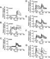
Mutant R205A disrupts PAR1-induced calcium mobilization in PAR1−/− astrocytes. (A) Vector only (control), (B) wtPAR1, or the indicated (C–G) PAR1 i2 loop mutant cDNA was nucleofected into astrocytes harvested from PAR1−/− mice. Two days later, the cells were loaded with Fura-2 for 30–40 min, and coverslips were transferred to a microscope stage for imaging. Imaging was performed with dual excitation at 340 nm and 380 nm wavelengths and the two resulting images were used for ratio calculations (Ex 340/Ex 380). Individual traces from 5 separate experiments for each receptor are shown superimposed on the same graph.
Figure 5
Figure 5
Mutant R205A disrupts PAR1-stimulated InsP signaling in PAR1−/− astrocytes. Astrocytes from mice lacking the PAR1 gene (PAR1−/−) were harvested and nucleofected with either plasmid vector alone (control), wild type PAR1-mCherry, or the PAR1-R205A i2 loop mutant cDNA. Two days after the nucleofection period, cells were or incubated in either serum-free media overnight (A–B) or this media containing 10 µCi/mL myo-[3H]inositol (C). The next day, cells were either fixed for confocal imaging (A–B) or incubated with LiCl2 and then stimulated with 50 µM TFLLR for 30 min (C). For the measurement of inositol phosphates (C), reactions were stopped by solubilizing astrocytes in formic acid. After samples were subjected to anion exchange chromatography, total [3H]InsPs were measured using liquid scintillation spectrometry. The accumulation of [3H]InsP is shown as counts per minute from a representative experiment (data are pooled results, mean cpm +/− S.E.M, from n=2 experiments, each performed in tripiclate).
Figure 6
Figure 6
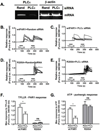
PLCε siRNA knock-down interferes with calcium mobilization in PAR1−/− astrocytes. A) Astrocytes harvested from PAR1−/− mice were treated with 100 MOI PLCε or random siRNA for 4 hr. Efficiency of knock-down was determined by RT-PCR three days after transduction. B–E) For calcium imaging experiments, PAR1−/− astrocytes were nucleofected with wild-type (wt) PAR1 or PAR1-R205A cDNA. Two days later, the cells were treated with 100 MOI PLCε or random siRNA. Following an incubation of three additional days, the astrocytes were loaded with Fura-2 for 30–40 min, and coverslips were transferred to a microscope stage for imaging. Imaging was performed with dual excitation at 340 nm and 380 nm wavelengths and the two resulting images were used for ratio calculations (Ex 340/Ex 380). The ratio at each time point was normalized to the average ratio before stimulation with 50 µM TFLLR. F, G) Quantification of calcium responses. The maximum responses to TFLLR (F) and ATP (G) were determined for each cell and averaged across each group.

Similar articles

Cited by

References

    1. Vu TK, Hung DT, Wheaton VI, Coughlin SR. Cell. 1991;64:1057–1068. - PubMed
    1. Coughlin SR. J Thromb.Haemost. 2005;3:1800–1814. - PubMed
    1. Gingrich MB, Traynelis SF. Trends in Neurosciences. 2000;23:399–407. - PubMed
    1. Ossovskaya VS, Bunnett NW. Physiol Rev. 2004;84:579–621. - PubMed
    1. Traynelis SF, Trejo J. Curr.Opin.Hematol. 2007;14:230–235. - PubMed

Publication types

MeSH terms

Substances

LinkOut - more resources