Clinical Trial
. 2012 Feb;9(1):016004.
doi: 10.1088/1478-3975/9/1/016004. Epub 2012 Feb 3.

High-definition imaging of circulating tumor cells and associated cellular events in non-small cell lung cancer patients: a longitudinal analysis

Affiliations
Clinical Trial

High-definition imaging of circulating tumor cells and associated cellular events in non-small cell lung cancer patients: a longitudinal analysis

Jorge Nieva et al. Phys Biol. 2012 Feb.

Abstract

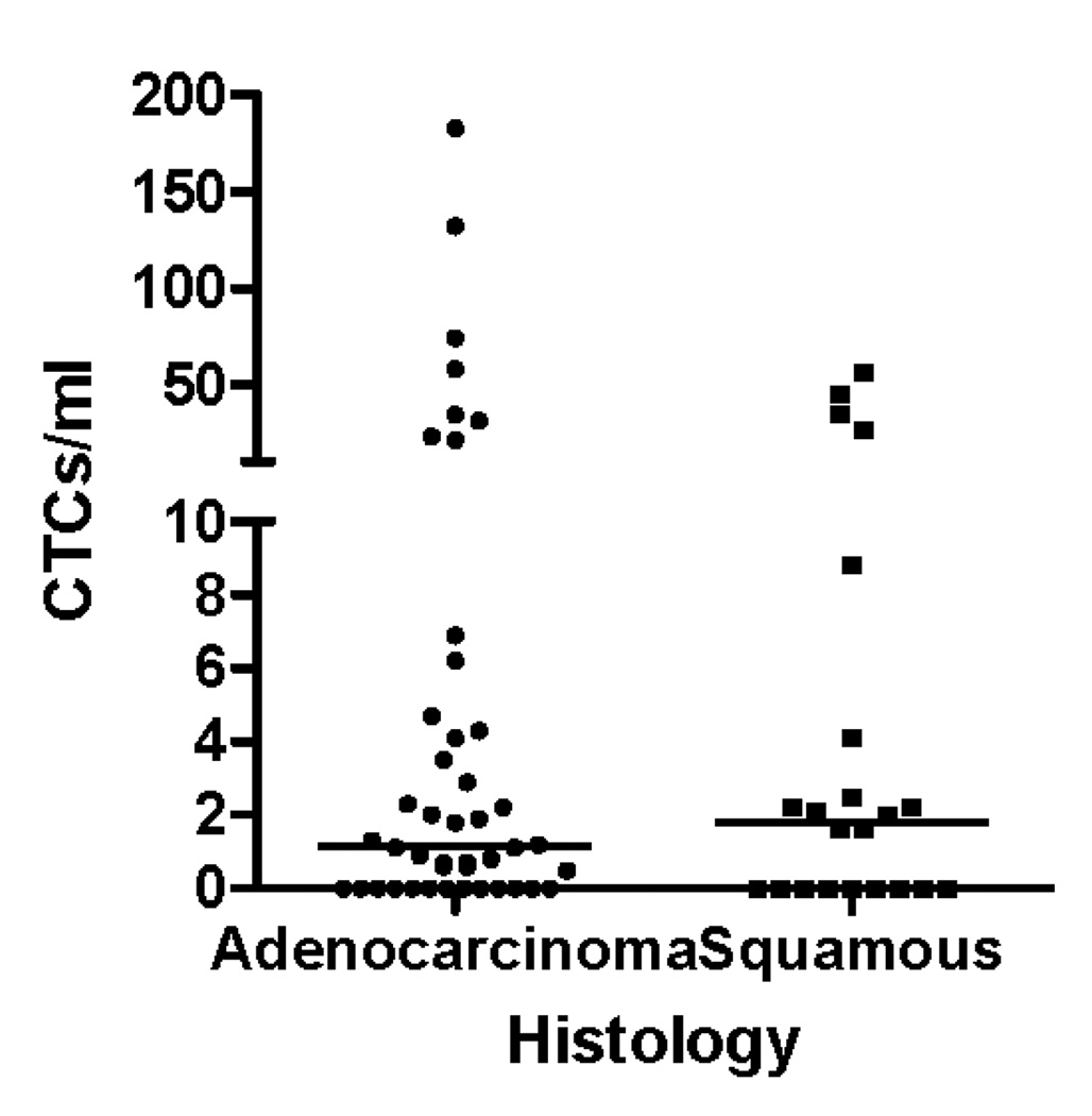
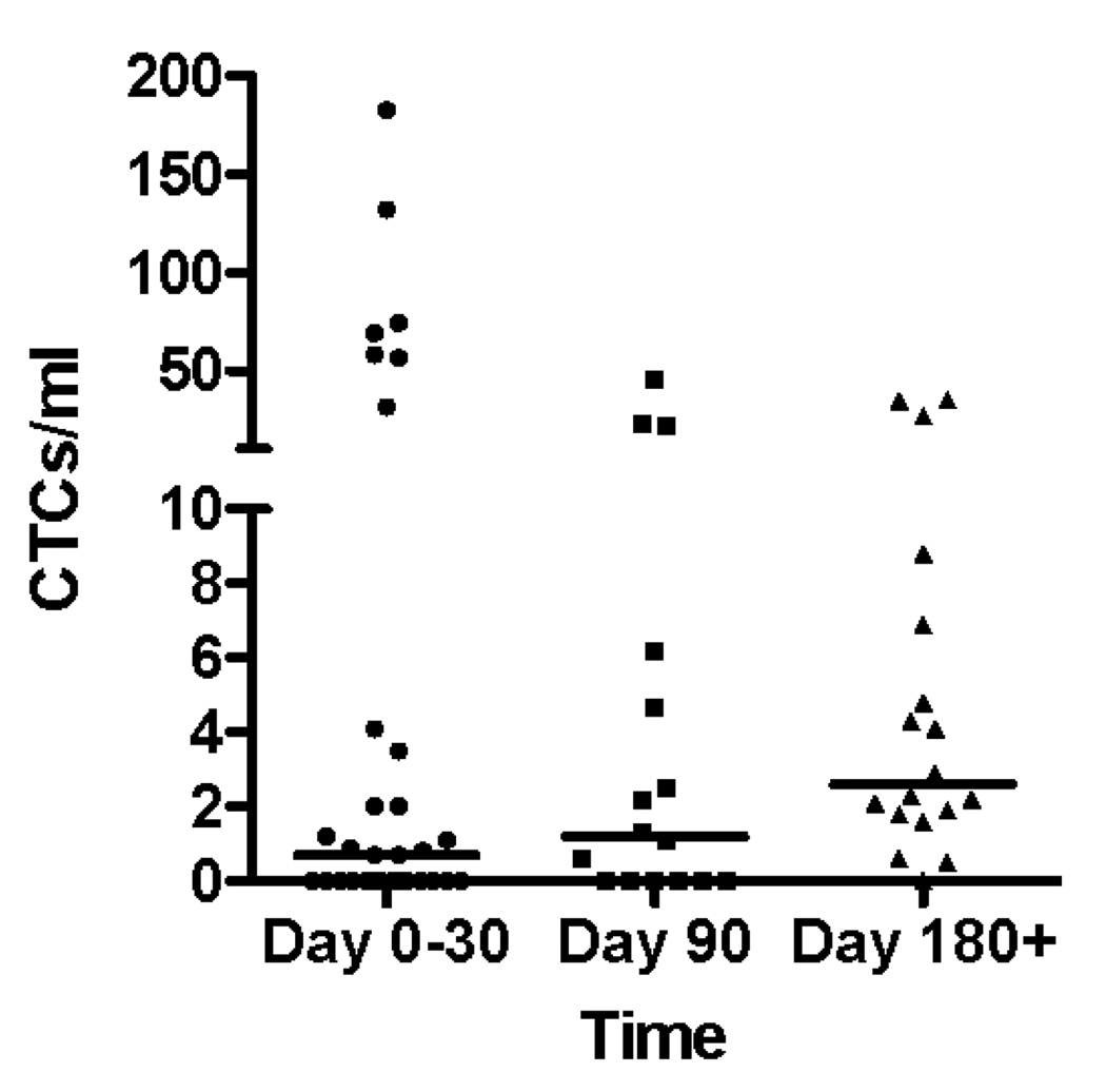
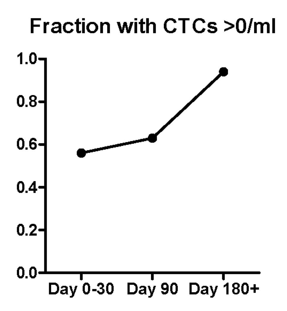
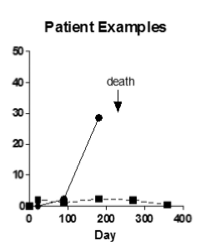
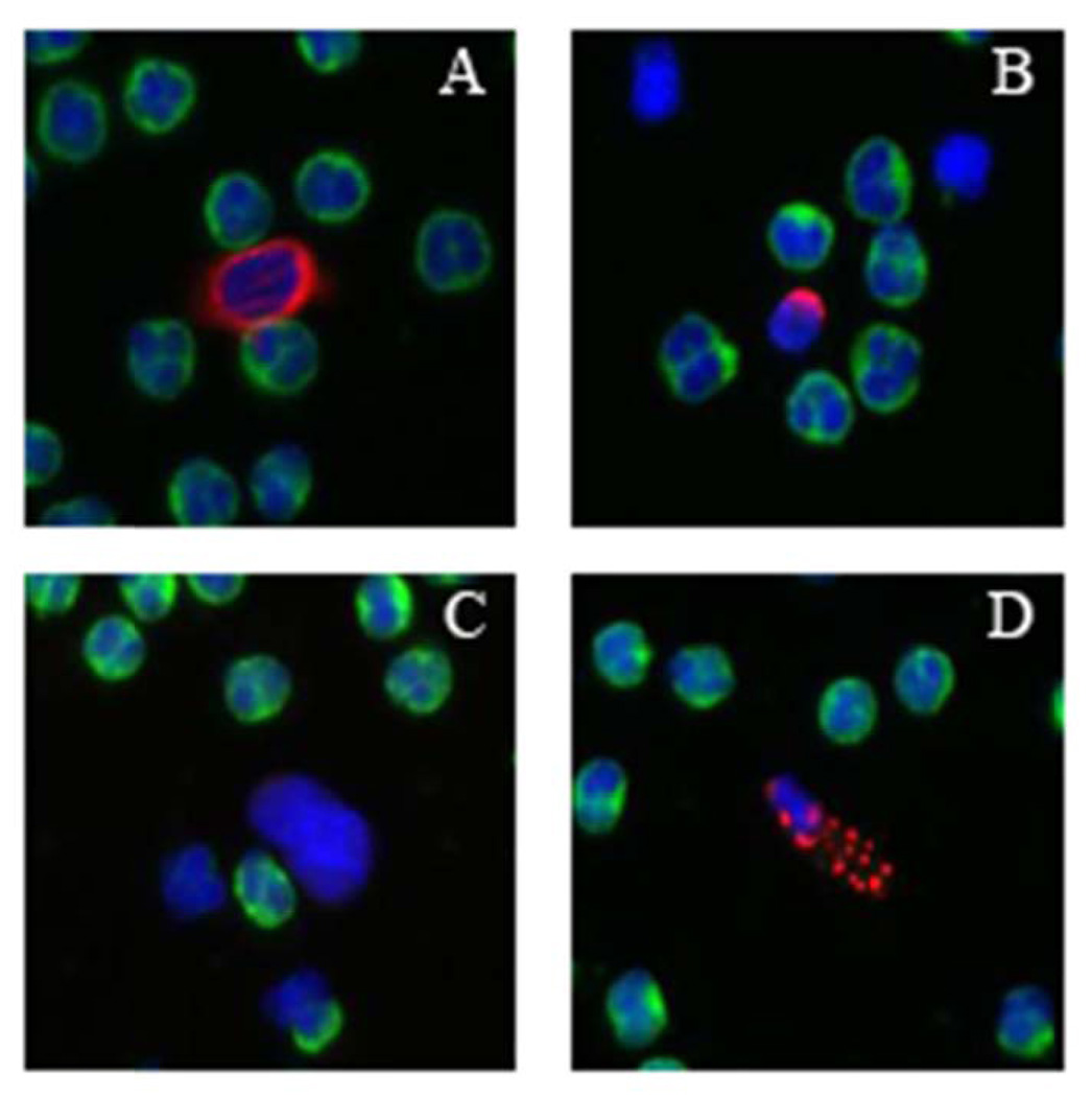
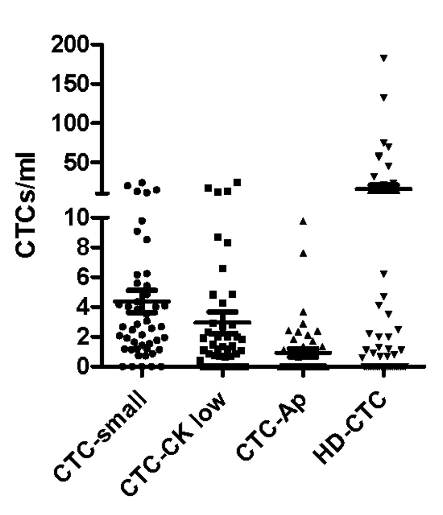
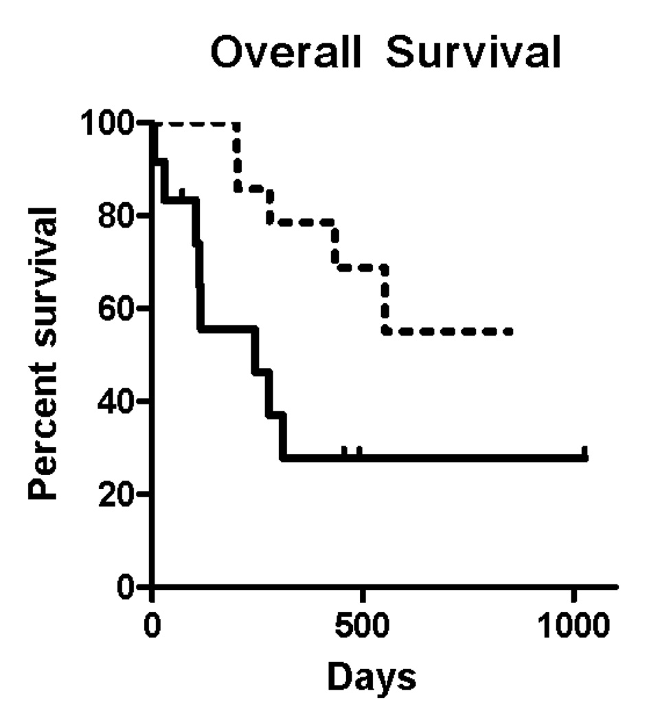
Sampling circulating tumor cells (CTCs) from peripheral blood is ideally accomplished using assays that detect high numbers of cells and preserve them for downstream characterization. We sought to evaluate a method using enrichment free fluorescent labeling of CTCs followed by automated digital microscopy in patients with non-small cell lung cancer. Twenty-eight patients with non-small cell lung cancer and hematogenously seeded metastasis were analyzed with multiple blood draws. We detected CTCs in 68% of analyzed samples and found a propensity for increased CTC detection as the disease progressed in individual patients. CTCs were present at a median concentration of 1.6 CTCs ml⁻¹ of analyzed blood in the patient population. Higher numbers of detected CTCs were associated with an unfavorable prognosis.


Figures

Figure 1
Figure 1
The fluid biopsy assay begins with patient blood tubes and the removal of red blood cells (RBCs) via lysis and then plating the remaining cells onto a slide. After antibody incubation, the full active surface of each slide is imaged in 3 fluorescence channels. Image analysis is used to extract features and associated intensity and cytomorphological values for each cell and the data is reduced for evaluation by a technical analyst.
Figure 2
Figure 2
(A) CTC counts from samples from adenocarcinoma and squamous carcinoma patients do not differ according to histologic subtype of lung cancer. (B) CTC counts for the whole patient population at early, middle and late timepoints with bar representing the median CTC count at the 3 time points. (C) during the natural history of stage IV NSCLC at early, middle, and late timepoints the frequency with which patients had a non-zero value for the CTC count appeared to increase. (D) Examples of serial blood draws on two patient types. The solid line represents a patient who had undetectable CTCs early in the course of the disease, then progressed and died 45 days after the last sample showed an increase in the CTC count. The dotted line represents a patient who maintained low CTC counts and remained alive at last follow-up 345 days after the 1 year timepoint of the study.
Figure 2
Figure 2
(A) CTC counts from samples from adenocarcinoma and squamous carcinoma patients do not differ according to histologic subtype of lung cancer. (B) CTC counts for the whole patient population at early, middle and late timepoints with bar representing the median CTC count at the 3 time points. (C) during the natural history of stage IV NSCLC at early, middle, and late timepoints the frequency with which patients had a non-zero value for the CTC count appeared to increase. (D) Examples of serial blood draws on two patient types. The solid line represents a patient who had undetectable CTCs early in the course of the disease, then progressed and died 45 days after the last sample showed an increase in the CTC count. The dotted line represents a patient who maintained low CTC counts and remained alive at last follow-up 345 days after the 1 year timepoint of the study.
Figure 3
Figure 3
In addition to cells that are morphologically consistent with CTCs, other related cells are also detected by the assay system and are observed in lung cancer patients. (A) CTC (B) CTC-small (C) CTC-CK low (D) CTC-Ap (E) counts of the different related cells in NSCLC population of patients
Figure 3
Figure 3
In addition to cells that are morphologically consistent with CTCs, other related cells are also detected by the assay system and are observed in lung cancer patients. (A) CTC (B) CTC-small (C) CTC-CK low (D) CTC-Ap (E) counts of the different related cells in NSCLC population of patients
Figure 4
Figure 4
(A) Overall survival for the entire study population based on averaging all CTC counts for each individual patients and grouping them into patients with 5 or more (solid line) and fewer than 5 CTCs/ml (dotted line). Hazard ratio for death was 4.0 (95% CI 1.242 to 13.14) comparing the group with high versus low CTC group. (B) For the subgroup of patient who died while enrolled in the study, the time in days from measurement of CTCs until death for 28 blood specimens obtained from 13 patients was recorded. The detection of 5 or more CTCs/mL was associated with a shorter period of time before death occurred, as compared with those samples where 5 or fewer CTCs/mL were observed.
Figure 4
Figure 4
(A) Overall survival for the entire study population based on averaging all CTC counts for each individual patients and grouping them into patients with 5 or more (solid line) and fewer than 5 CTCs/ml (dotted line). Hazard ratio for death was 4.0 (95% CI 1.242 to 13.14) comparing the group with high versus low CTC group. (B) For the subgroup of patient who died while enrolled in the study, the time in days from measurement of CTCs until death for 28 blood specimens obtained from 13 patients was recorded. The detection of 5 or more CTCs/mL was associated with a shorter period of time before death occurred, as compared with those samples where 5 or fewer CTCs/mL were observed.

References

    1. Cristofanilli M, Budd GT, Ellis MJ, Stopeck A, Matera J, Miller MC, Reuben JM, Doyle GV, Allard WJ, Terstappen LW, Hayes DF. Circulating tumor cells, disease progression, and survival in metastatic breast cancer. N. Engl. J. Med. 2004;351:781–791. - PubMed
    1. Sastre J, Maestro ML, Puente J, Veganzones S, Alfonso R, Rafael S, García-Saenz JA, Vidaurreta M, Martíin M, Arroyo M, Sanz-Casla MT, Díaz-Rubio E. Circulating tumor cells in colorectal cancer: correlation with clinical and pathological variables. Ann. of Oncol. 2008;19:935–938. - PubMed
    1. Ghossein RA, Scher HI, Gerald WL, Kelly WK, Curely T, Amsterdam A, Zhang ZF, Rosai J. Detection of circulating tumor cells in patients with localized and metastatic prostatic carcinoma: clinical implications. J. Clin. Oncol. 1995;13:1195–1200. - PubMed
    1. Krebs MG, Sloane R, Priest L, Lancashire L, Hou JM, Greystoke A, Ward TH, Ferraldeschi R, Hughes A, Clack G, Ranson M, Dive C, Blackhall FH. Evaluation and prognostic significance of circulating tumor cells in patients with non-small-cell lung cancer. J. Clin. Oncol. 2011;29:1556–1563. - PubMed
    1. Gervasoni A, Sandri MT, Nascimbeni R, Zorzino L, Cassatella MC, Baglioni L, Panigara S, Gervasi M, Di Lorenzo D, Parolini O. Comparison of three distinct methods for the detection of circulating tumor cells in colorectal cancer patients. Oncol. Rep. 2011;25:1669–1703. - PubMed

Publication types

MeSH terms