Review
. 2012 Mar;9(2):113-22.
doi: 10.1038/cmi.2011.60. Epub 2012 Feb 6.

Peli: a family of signal-responsive E3 ubiquitin ligases mediating TLR signaling and T-cell tolerance

Affiliations
Review

Peli: a family of signal-responsive E3 ubiquitin ligases mediating TLR signaling and T-cell tolerance

Wei Jin et al. Cell Mol Immunol. 2012 Mar.

Abstract

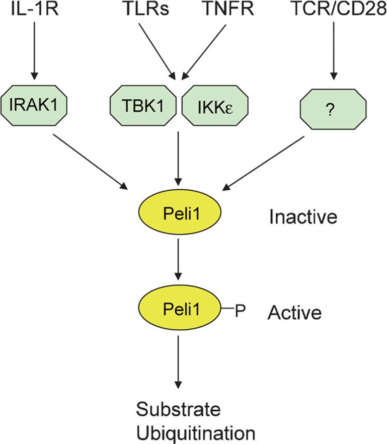
E3 ubiquitin ligases play a crucial role in regulating immune receptor signaling and modulating immune homeostasis and activation. One emerging family of such E3s is the Pelle-interacting (Peli) proteins, characterized by the presence of a cryptic forkhead-associated domain involved in substrate binding and an atypical RING domain mediating formation of both lysine (K) 63- and K48-linked polyubiquitin chains. A well-recognized function of Peli family members is participation in the signal transduction mediated by Toll-like receptors (TLRs) and IL-1 receptor. Recent gene targeting studies have provided important insights into the in vivo functions of Peli1 in the regulation of TLR signaling and inflammation. These studies have also extended the biological functions of Peli1 to the regulation of T-cell tolerance. Consistent with its immunoregulatory functions, Peli1 responds to different immune stimuli for its gene expression and catalytic activation. In this review, we discuss the recent progress, as well as the historical perspectives in the regulation and biological functions of Peli.


Figures

Figure 1
Figure 1
Signal-induced Peli1 activation. Peli1 exists as an inactive form but becomes activated in response to signals from different immune receptors. Peli1 activation involves its phosphorylation by IRAK1 and the IKK-related kinases TBK1 and IKKε. Whereas IRAK1 mediates Peli1 activation by IL-1R, TBK1 and IKKε mediate Peli1 activation by TLRs and TNFR. It remains unclear how Peli1 is activated in T cells by the TCR and CD28 signals. IKK, IκB kinase; IRAK, IL-1R-associated kinase; Peli1, Pelle-interacting protein 1; TCR, T-cell receptor; TLR, Toll-like receptor; TNFR, tumor-necrosis factor receptor.
Figure 2
Figure 2
Peli1 mediates IKK activation by the TRIF-dependent TLR pathway. Based on the signaling adaptors, TLR signaling can be classified into two major pathways: the MyD88- and TRIF-dependent pathways. TLR3 is the only TLR that exclusively signals through TRIF, whereas TLR4 signals through both TRIF and MyD88. The rest of the TLRs, as well as IL-1R, signal through MyD88. An important mechanism of TLR signaling is K63 type ubiquitination of signaling adaptors, including TRAF6, RIP1 and TRAF3, which mediate activation of downstream signaling events. Activation of IKK/NF-κB by the TRIF-dependent TLR pathway, but not that of the MyD88 pathway, depends on Peli1. Peli1 functions by mediating ubiquitination of RIP1 and, thereby, activating IKK and NF-κB. The TRAF3-regulated signaling pathway may crosstalk with the RIP1 pathway through induction of Peli1 gene expression and TBK1-/IKKε-mediated Peli1 activation. IKK, IκB kinase; Peli1, Pelle-interacting protein 1; RIP1, receptor-interacting protein 1; TCR, T-cell receptor; TLR, Toll-like receptor.
Figure 3
Figure 3
Peli1 negatively regulates T-cell activation by mediating c-Rel degradation. In T cells, c-Rel is an important NF-κB member that regulates IL-2 gene expression and T-cell activation. c-Rel functions as either an homodimer or heterodimers associated with different NF-κB members, such as p50 and RelA. Upon IKK-mediated degradation of IκB, the c-Rel-NF-κB dimers translocate to the nucleus, where they participate in the transactivation of IL-2 and other target genes. The TCR/CD28 signals also stimulate the expression of Peli1 via a yet-to-be-characterized transcription factor(s), and the accumulated Peli1 negatively regulates the late-phase activation of NF-κB by mediating ubiquitination and degradation of c-Rel. It is currently unknown whether Peli1 exerts this function in the cytoplasm or nucleus, but Peli1 is located in both of these cellular compartments (Chang M et al., unpubl. data). IKK, IκB kinase; Peli1, Pelle-interacting protein 1; RIP1, receptor-interacting protein 1; TCR, T-cell receptor; TLR, Toll-like receptor.

References

    1. Hershko A, Ciechanover A. The ubiquitin system. Annu Rev Biochem. 1998;67:425–479. - PubMed
    1. Chen ZJ, Sun LJ. Nonproteolytic functions of ubiquitin in cell signaling. Mol Cell. 2009;33:275–286. - PubMed
    1. Sun SC. Deubiquitylation and regulation of the immune response. Nat Rev Immunol. 2008;8:501–511. - PMC - PubMed
    1. Ardley HC, Robinson PA. E3 ubiquitin ligases. Essays Biochem. 2005;41:15–30. - PubMed
    1. Li W, Bengtson MH, Ulbrich A, Matsuda A, Reddy VA, Orth A, et al. Genome-wide and functional annotation of human E3 ubiquitin ligases identifies MULAN, a mitochondrial E3 that regulates the organelle's dynamics and signaling. PLoS ONE. 2008;3:e1487. - PMC - PubMed

Publication types