Review
. 2012 Mar;96(3):304-21.
doi: 10.1016/j.pneurobio.2012.01.009. Epub 2012 Jan 28.

GPCR mediated regulation of synaptic transmission

Affiliations
Review

GPCR mediated regulation of synaptic transmission

Katherine M Betke et al. Prog Neurobiol. 2012 Mar.

Abstract

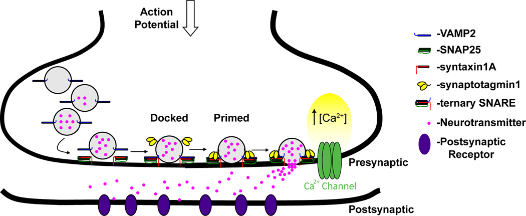
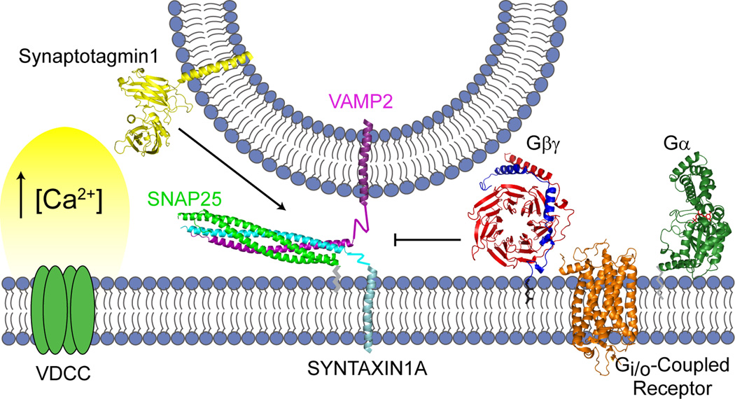
Synaptic transmission is a finely regulated mechanism of neuronal communication. The release of neurotransmitter at the synapse is not only the reflection of membrane depolarization events, but rather, is the summation of interactions between ion channels, G protein coupled receptors, second messengers, and the exocytotic machinery itself which exposes the components within a synaptic vesicle to the synaptic cleft. The focus of this review is to explore the role of G protein signaling as it relates to neurotransmission, as well as to discuss the recently determined inhibitory mechanism of Gβγ dimers acting directly on the exocytotic machinery proteins to inhibit neurotransmitter release.


Figures

Figure 1
Figure 1. Overview of neurotransmission across a synapse
In the presynaptic neuron, synaptic vesicles expressing VAMP2, among other proteins embedded in their membrane, are loaded with neurotransmitters. Simplistically, docking of the vesicle occurs via interaction of VAMP2 with t-SNARE (SNAP-25/syntaxin 1A), a process that is guided and regulated by numerous proteins, while priming has been described as further zippering of the SNARE complex with interaction of other major components such as synaptotagmin, MUNC13, and MUNC18. Upon the arrival of an action potential, VDCC facilitate a large increase in intracellular calcium concentration. This rise is sensed by proteins within the synaptic complex of the vesicle at the membrane such that fusion of the vesicle membrane with the presynaptic membrane occurs, resulting in release of neurotransmitter into the synaptic cleft to activate its respective receptor on the post-synaptic neuron ensuring propagation of action potentials from one neuron to another.
Figure 2
Figure 2. Structure of G protein Coupled Receptors and Heterotrimeric G proteins
Shown in ribbon diagrams are representative examples of GPCRs and heterotrimeric G proteins. A) The structure of rhodopsin (PDB ID: 1F88) (Palczewski et al., 2000) is shown in orange, and it depicts the seven transmembrane α-helices and the respective extracellular and intracellular surfaces that interact with agonists and heterotrimeric G proteins, respectively. B) The structure of the heterotrimer transducin (PDB ID: 1GOT) (Lambright et al., 1996) is shown. The Gα subunit (green) has two domains, a GTPase domain and an α-helical domain. Within the GTPase domain, there are three regions termed Switch I, II, and III, (C), that have different orientations depending on which guanine nucleotide is present. In the GDP-bound form, Switches I–III form the major interface that interacts with the Gβγ dimer. The structure of the Gβ subunit (red) is an N-terminal α-helix followed by a series of β sheets that make a seven-bladed propeller or toroid. A single blade (light blue) is based on the amino acid motif termed WD-40 repeat. The structure of the Gγ subunit (dark blue) is two tandem α-helices that form interactions with the N-terminal α-helix of Gβ and a surface of the Gβ toroid on the opposite face from where Gα interacts with Gβ.
Figure 3
Figure 3. The interactions with Gβγ during exocytosis
Gβγ subunits are known to have a variety of interactions during exocytosis - GPCRs and Gα subunits during its activation, as well as a number of effectors. Shown are the structures, if known, of those proteins that interact with Gβγ and play a role in exocytosis. THE GPCRs and Gα are discussed in Figure 2. The SNARE proteins (SNAP-25/Syntaxin 1A/VAMP2) form a trimeric complex through interaction of their α-helical SNARE motifs, SNAP-25 having 2 two of them (PDB ID: 1SFC)(Sutton et al., 1998). VAMP2 (purple) and syntaxin 1A (cyan) have transmembrane domains through which they interact with membranes. SNAP-25 (green) has a palmitoylation site near the C-terminal portion of its first SNARE motif by which it is tethered to the membrane. Not shown is the N-terminal helical domain of syntaxin 1A, Habc. The crystal structure of syntaxin 1A without the transmembrane domain confirms the triple helix Habc domain at its N-terminus. The GIRK channel is represented by the crystal structure for the structurally similar ATP-sensitive Kir channel 10 which is a tetramer with both transmembrane and intracellular domains (PDB ID: 2X6A) (Clarke et al., 2010). There is no structure determined yet for VDCC; it is shown as a simple transmembrane cartoon.
Figure 4
Figure 4. Hypothesis of Gβγ regulation of presynaptic vesicle release
Synaptic vesicles are primed by a tethering interaction between VAMP2 on the vesicle and the SNAP-25/syntaxin 1A dimer at the plasma membrane. At low intracellular concentrations of calcium, activation of Gi/o-coupled receptors results in release of Gβγ that will bind to the SNARE proteins and prevent binding of synaptotagmin. However, high enough intracellular calcium concentrations, such as with repetitive neuronal stimulation, synaptotagmin is able to compete with Gβγ for binding to SNARE, and thereby promote fusion of the vesicles with the plasma membrane. Figure adapted from Wells et al. (Wells et al., 2011).

Similar articles

Cited by

References

    1. Ahnert-Hilger G, Brautigam M, Gratzl M. Ca2+-stimulated catecholamine release from alpha-toxin-permeabilized PC12 cells: biochemical evidence for exocytosis and its modulation by protein kinase C and G proteins. Biochemistry. 1987;26(24):7842–7848. - PubMed
    1. Albert PR, Robillard L. G protein specificity: traffic direction required. Cell. Signal. 2002;14(5):407–418. - PubMed
    1. Albrecht C, Bloss HG, Jackisch R, Feuerstein TJ. Evaluation of autoreceptor-mediated control of [3H]acetylcholine release in rat and human neocortex. Exp. Brain Res. 1999;128(3):383–389. - PubMed
    1. Albsoul-Younes AM, Sternweis PM, Zhao P, Nakata H, Nakajima S, Nakajima Y, Kozasa T. Interaction sites of the G protein beta subunit with brain G protein-coupled inward rectifier K+ channel. J. Biol. Chem. 2001;276(16):12712–12717. - PubMed
    1. Allen TG, Brown DA. M2 muscarinic receptor-mediated inhibition of the Ca2+ current in rat magnocellular cholinergic basal forebrain neurones. J. Physiol. 1993;466:173–189. - PMC - PubMed

Publication types

MeSH terms

Substances