. 2012 Apr;78(8):2522-32.
doi: 10.1128/AEM.06252-11. Epub 2012 Feb 3.

Equine stomachs harbor an abundant and diverse mucosal microbiota

Affiliations

Equine stomachs harbor an abundant and diverse mucosal microbiota

G A Perkins et al. Appl Environ Microbiol. 2012 Apr.

Abstract

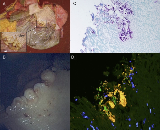
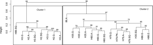
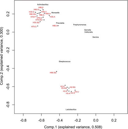
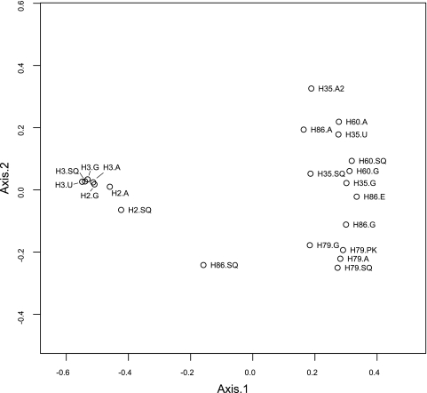
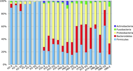
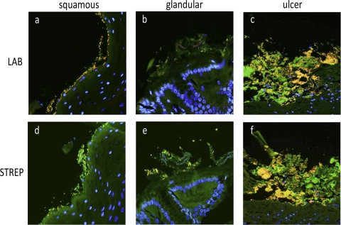
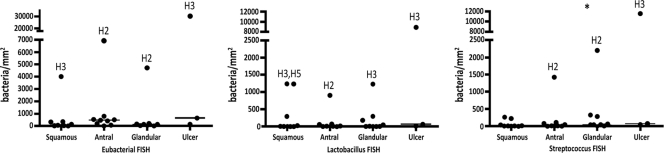
Little is known about the gastric mucosal microbiota in healthy horses, and its role in gastric disease has not been critically examined. The present study used a combination of 16S rRNA bacterial tag-encoded pyrosequencing (bTEFAP) and fluorescence in situ hybridization (FISH) to characterize the composition and spatial distribution of selected gastric mucosal microbiota of healthy horses. Biopsy specimens of the squamous, glandular, antral, and any ulcerated mucosa were obtained from 6 healthy horses by gastroscopy and from 3 horses immediately postmortem. Pyrosequencing was performed on biopsy specimens from 6 of the horses and yielded 53,920 reads in total, with 631 to 4,345 reads in each region per horse. The microbiome segregated into two distinct clusters comprised of horses that were stabled, fed hay, and sampled at postmortem (cluster 1) and horses that were pastured on grass, fed hay, and biopsied gastroscopically after a 12-h fast (cluster 2). The types of bacteria obtained from different anatomic regions clustered by horse rather than region. The dominant bacteria in cluster 1 were Firmicutes (>83% reads/sample), mainly Streptococcus spp., Lactobacillus spp. and, Sarcina spp. Cluster 2 was more diverse, with predominantly Proteobacteria, Bacteroidetes, and Firmicutes, consisting of Actinobacillus spp. Moraxella spp., Prevotella spp., and Porphyromonas spp. Helicobacter sp. sequences were not identified in any of 53,920 reads. FISH (n = 9) revealed bacteria throughout the stomach in close apposition to the mucosa, with significantly more Streptococcus spp. present in the glandular region of the stomach. The equine stomach harbors an abundant and diverse mucosal microbiota that varies by individual.


Figures

Fig 1
Fig 1
The equine stomach during gross postmortem and endoscopic examinations followed by a closer investigation with histopathology and FISH. (A) Postmortem examination of H3, with a grade 2 ulcer in the squamous portion of the stomach near the margo plicatus. (B) Endoscopic view of the cardiac region (H158) of the stomach, with small pinpoint ulcers (grade 1) at the junction of the squamous and glandular regions. (C) Gram stain of ulcerated mucosa (H158) shows colonization by Gram-positive cocci and rods (magnification, ×40). (D) FISH with probes directed against bacteria in general (EUB338, 6-FAM) and Streptococcus spp. (STREP, Cy-3), showing dense colonization of the ulcerated mucosa by Streptococcus spp. (bright yellow) and fewer other bacteria (green). DAPI (4′,6′-diamidino-2-phenylindole)-stained nuclei appear blue. Original magnification, ×60.
Fig 2
Fig 2
Hierarchical cluster analysis of samples of the equine gastric mucosa based on relative abundance of sequence reads on the bacterial genus level. The two main clusters found in this analysis coincide with differences in management (feeding, exercise, housing, etc.) and sampling method. Horses sampled immediately postmortem (H2 and H3) and fed hay formed cluster 1, whereas horses biopsied endoscopically after a 12-hour fast (H35, H60, and H79) and fed hay and grass formed cluster 2. The exception was horse H86, which appeared in both clusters. An individual horse had a distinct equine gastric mucosa microbiota without differences between regions. Numbers above the nodes are approximate unbiased values. Labels indicate horse identification number along with stomach region (Sq, squamous; G, glandular; A, antral; U, ulcer; E, erosion; PK, superficial parakeratosis).
Fig 3
Fig 3
Principal-component analysis plot of the horse stomach samples (dots) based on the relative abundance of bacterial genera per horse. Labels indicate horse identification number and stomach region (SQ, squamous; G, glandular; A, antral; U, ulcer; E, erosion; P, superficial parakeratosis). Names of the bacterial genera with the highest loadings have been placed in the plot according to their correlation to the first two components.
Fig 4
Fig 4
Principal-coordinate ordination plot of the samples from the horses, based on a Bray-Curtis matrix derived from the relative abundance of bacterial genera per horse. Labels indicate horse identification number and stomach region (SQ, squamous; G, glandular; A, antral; U, ulcer; E, erosion; PK, = superficial parakeratosis).
Fig 5
Fig 5
Top five phyla found in the gastric mucosae of 6 healthy horses by bTEFAP. The top five phyla represented were Firmicutes, Bacteroidetes, Proteobacteria, Fusobacteria, and Actinobacteria. Cluster 1 horses (H2 and H3) had less diversity, with a predominance of Firmicutes (>82%), whereas horses in cluster 2 (H35, H60, H79, and H86) had a more diverse population consisting of higher percentages of Proteobacteria and Bacteroidetes, as well as Firmicutes. The horse identification number and stomach region (SQ, squamous; G, glandular; A, antral; U, ulcer; E, erosion; PK, superficial parakeratosis) are along the x axis.
Fig 6
Fig 6
FISH shows abundant bacteria in close apposition to the equine gastric mucosa. Histologic sections from the squamous (a and d), glandular (b and e), and ulcerated (c and f) mucosa were examined using labeled oligonucleotide probes directed against bacteria in general (EUB-338, 6-FAM, green) in combination with probes directed against Lactobacillus spp. (a, b, and c) (Cy-3) or Streptococcus spp. (d, e, and f) (Cy-3). In healthy squamous mucosa, Lactobacillus spp. (a) (yellow) and Streptococcus spp. (d) (yellow) are in close apposition to the epithelium. In the glandular region, both Lactobacillus spp. (b) (yellow) and Streptococcus spp. (e) (yellow) colonize the mucosa but appear to be present in smaller amounts than in the squamous region. Ulcerated mucosa from H3 (c and f) contains an abundant mixed bacterial population that includes Streptococcus spp. (f) (yellow), Lactobacillus spp. (c) (yellow), and other bacterial species (c and f) (green). Nuclei are blue (DAPI). Original magnification, ×600.
Fig 7
Fig 7
Healthy horses generally have an abundance of bacteria of <1,000 bacteria/mm2 in all gastric mucosal samples by as determined by FISH (n = 9). FISH was done with probes directed against bacteria in general (eubacteria), Streptococcus spp., and Lactobacillus spp. A few horses (H2, H3, and H35) had outliers as indicated. Horse 3's grade 2 ulcer had an abundance of bacteria (30,529 bacteria/mm2). All other horses had small pinpoint ulcers, with total numbers of bacteria similar to those in other regions of the stomach. Each symbol represents a horse. There were significantly more Streptococcus bacteria in the glandular mucosa than in the squamous and antral mucosae (P = 0.0098), as indicated by the asterisk.

Similar articles

Cited by

References

    1. Acosta-Martinez V, Dowd SE, Sun Y, Allen V. 2009. Tag-encoded pyrosequencing analysis of bacterial diversity in a single soil type as affected by management and land use. Soil Biol. Biochem. 4:2762–2770
    1. Alexander F. 1972. Certain aspects of the physiology and pharmacology of the horse's digestive tract. Equine Vet. J. 4:166–169
    1. Al Jassim RAM, Scott T, Trebbin AL, Trott D, Pollitt CC. 2005. The genetic diversity of lactic acid producing bacteria in the equine gastrointestinal tract. FEMS Microbiol. Lett. 248:75–81 - PubMed
    1. Al Jassim RAM. 2006. Supplementary feeding of horses with processed sorghum grains and oats. Anim. Feed Sci. Tech. 125:33–44
    1. Andrews FM, Buchanan BR, Elliot SB, Clairday NA, Edwards LH. 2005. Gastric ulcers in horses. J. Anim. Sci. 83:E18–E21

Publication types

LinkOut - more resources