. 2012 Apr;78(8):2841-9.
doi: 10.1128/AEM.07274-11. Epub 2012 Feb 3.

Pathogenicity and infection cycle of Vibrio owensii in larviculture of the ornate spiny lobster (Panulirus ornatus)

Affiliations

Pathogenicity and infection cycle of Vibrio owensii in larviculture of the ornate spiny lobster (Panulirus ornatus)

Evan F Goulden et al. Appl Environ Microbiol. 2012 Apr.

Abstract

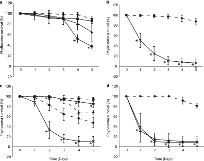
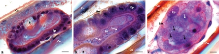
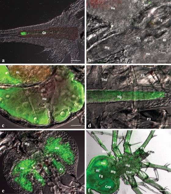
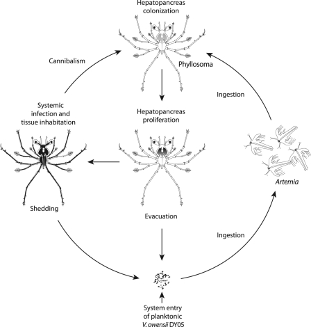
The type strain of Vibrio owensii (DY05) was isolated during an epizootic of aquaculture-reared larvae (phyllosomas) of the ornate spiny lobster (Panulirus ornatus). V. owensii DY05 was formally demonstrated to be the etiological agent of a disease causing rapid and reproducible larval mortality with pathologies similar to those seen during disease epizootics. Vectored challenge via the aquaculture live feed organism Artemia (brine shrimp) caused consistent cumulative mortality rates of 84 to 89% after 72 h, in contrast to variable mortality rates seen after immersion challenge. Histopathological examination of vector-challenged phyllosomas revealed bacterial proliferation in the midgut gland (hepatopancreas) concomitant with epithelial cell necrosis. A fluorescent-protein-labeled V. owensii DY05 transconjugant showed dispersal of single cells in the foregut and hepatopancreas 6 h postexposure, leading to colonization of the entire hepatopancreas within 18 h and eventually systemic infection. V. owensii DY05 is a marine enteropathogen highly virulent to P. ornatus phyllosoma that uses vector-mediated transmission and release from host association to a planktonic existence to perpetuate transfer. This understanding of the infection process will improve targeted biocontrol strategies and enhance the prospects of commercially viable larviculture for this valuable spiny lobster species.


Figures

Fig 1
Fig 1
Survival of P. ornatus phyllosomas experimentally infected with V. owensii DY05 by immersion (a) or vectored challenge via Artemia nauplii (b to d). (a) Stage 1 phyllosoma survival after immersion with V. owensii DY05 at concentrations of 0 (control; ●), 1 × 10³ (■), 1 × 10⁵ (▲), and 1 × 10⁷ (♦) CFU ml−1. Data from one of three independent experiments are presented, as high interexperimental variability prevented pooling of data. (b) Stage 1 phyllosoma survival after vectored challenge with nonenriched nauplii (control; ●) or nauplii enriched with V. owensii DY05 (×). Pooled data from three independent experiments are presented. (c) Stage 3 phyllosoma survival after vectored challenge with nonenriched nauplii (controls; ●, ■, and ▲) or nauplii enriched in V. owensii DY05 (×). Data from three independent experiments are shown; data for controls were not pooled due to high interexperimental variability, while data for V. owensii-challenged phyllosomas were pooled. The first experiment (control; ●) included a second control treated with an antibiotic cocktail (♦) prior to feeding with nauplii. (d) Stage 1 phyllosoma survival after vectored challenge with nonenriched nauplii (control; ●) or nauplii enriched with V. owensii DY05 (×) or DY05[GFP] (▲). The asterisks indicate data points significantly different (Student's t test) from the corresponding control. Means ± standard deviations (SD) are shown.
Fig 2
Fig 2
Histopathological analysis of stage 3 P. ornatus phyllosoma hepatopancreas tubules during vectored challenge with V. owensii DY05. (a) Distal end of tubule before exposure to V. owensii DY05, showing no visible bacterial cells, cuboidal epithelial cells, large lumen, and B-cell vacuoles. (b) Tubules 24 h after exposure, demonstrating bacterial proliferation (white arrows) within the lumen and rounding of epithelial cells. (c) Necrotic tubule 42 h after exposure, characterized by disintegration of epithelial cells (white arrow), cell sloughing, absence of defined lumen, and proliferation of bacteria (black arrows). B, B-cell vacuole; Bm, basement membrane; Ct, connective tissue; Cu, cuticle; Ep, epithelial cells; Lu, tubular lumen. All scale bars, 10 μm.
Fig 3
Fig 3
In situ spatiotemporal localization of V. owensii DY05[GFP] in Artemia nauplius after 2-h enrichment (a) and during the infection process of vector-challenged stage 1 P. ornatus phyllosomas (b to f). (a) Artemia nauplius showing colonization of DY05[GFP] in the gastrointestinal tract. Scale bar, 100 μm. (b) Hepatopancreas and midgut region of phyllosoma after 6-h exposure, demonstrating the presence of single cells or small aggregations of DY05[GFP] cells in the foregut, the midgut, and the anterior and lateral lobes of the hepatopancreas. Scale bar, 50 μm. (c) Proliferation of DY05[GFP] in the distal ends of the lateral and anterior hepatopancreas lobes 12 h after exposure. Scale bar, 50 μm. (d) Hindgut 12 h after exposure showing trafficking of DY05[GFP] cells followed by evacuation. Scale bar, 50 μm. (e) Hepatopancreas 18 h after exposure, showing illumination of the entire organ with fluorescent DY05[GFP] cells concomitant with tissue granulation and loss of architecture. Scale bar, 100 μm. (f) Dead phyllosoma 24 h after exposure, showing colonization of the entire body (systemic infection) by DY05[GFP] cells associated with loss of internal organ structural integrity. Scale bar, 200 μm. At, hepatopancreas anterior lobe; Cep, cephalic shield; Fg, foregut; Gt, gastrointestinal tract; He, hepatopancreas lateral lobe; Hg, hindgut; Mg, midgut; Mxp, maxilliped; P, pereiopod (1 to 3); Pln, pleon; Pv, pyloric valve; Thx, thorax.
Fig 4
Fig 4
Infection cycle of V. owensii DY05 in the larviculture ecosystem of early-stage P. ornatus phyllosomas. The pathogen enters the larviculture environment through an unascertained source as a free-living planktonic form. Live prey Artemia nauplii actively ingest the bacteria, resulting in bioaccumulation in the gut. Early-stage phyllosomas (stages 1 to 3) capture and masticate the Artemia nauplii, triturating food particulates along with the pathogen through the foregut into the hepatopancreas. The pathogen proliferates in the distal ends of the hepatopancreas, eventually colonizing the entire organ, with planktonic cells dispelled from the midgut and evacuated back into the ambient environment. These planktonic cells are ingested by Artemia nauplii, which perpetuates invasion of the phyllosomas through the oral route. Eventually, the condition progresses to systemic infection in association with acute mortality. By utilizing and inhabiting the tissues of moribund and dead hosts, the pathogen can also be transmitted to new hosts through occasional cannibalism. Continuous shedding of bacteria from dead hosts likely contributes to the ambient bacterioplankton, again reactivating the oral infection pathway.

Similar articles

Cited by

References

    1. Aguirre-Guzmán A, et al. 2010. Pathogenicity and infection route of Vibrio parahaemolyticus in American white shrimp, Litopenaeus vannamei. J. World Aquacult. Soc. 41:464–470
    1. Aguirre-Guzmán A, Vázquez-Juárez R, Ascencio F. 2001. Differences in the susceptibility of American white shrimp larval substages (Litopenaeus vannamei) to four Vibrio species. J. Invertebr. Pathol. 78:215–219 - PubMed
    1. Austin B, Zhang X-H. 2006. Vibrio harveyi: a significant pathogen of marine vertebrates and invertebrates. Lett. Appl. Microbiol. 43:119–124 - PubMed
    1. Austin B, Austin D, Sutherland R, Thompson F, Swings J. 2005. Pathogenicity of vibrios to rainbow trout (Oncorhynchus mykiss, Walbaum) and Artemia nauplii. Environ. Microbiol. 7:1488–1495 - PubMed
    1. Bourne DG, et al. 2004. Microbial community dynamics in a larval aquaculture system of the tropical rock lobster, Panulirus ornatus. Aquaculture 242:31–51

Publication types

LinkOut - more resources