Retrograde dynamic locked nailing for valgus knee correction: a revised technique
- PMID: 22307560
- PMCID: PMC3353075
- DOI: 10.1007/s00264-012-1495-8
Retrograde dynamic locked nailing for valgus knee correction: a revised technique
Abstract
Purpose: Traditionally, valgus knee deformity is predominately corrected by stabilisation with a plate inserted via the medial approach to the supracondylar region of the femur. However, this technique is unfavourable from both a biomechanical and a biological point of view. A revised retrograde dynamic locked nailing was developed to improve correction of this defect.
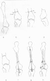
Method: Forty-one knees with valgus deformity (average tibiofemoral angle, 22°; range, 16-29°) in 25 adult patients were treated by oblique femoral supracondylar varus osteotomy and stabilised with retrograde dynamic locked nails. Postoperatively, early ambulation with protected weight bearing and range of motion knee exercises were encouraged.
Result: Thirty-five knees of 21 patients were followed-up for an average of 2.6 years (range, 1.1-4.5 years). All osteotomy sites healed with an average union period of 3.4 months (range, 2.5-5.0 months). There were no significant complications. At the latest follow-up, the average tibiofemoral angle was 7.1° valgus (range, 4-10° valgus). For all of the knees, the outcomes were satisfactory (p < 0.001).
Conclusion: The technique described here may be a feasible alternative for correction of valgus knee deformity. The advantages of this technique include the use of a biomechanically more appropriate method, a minimal complication rate and a high rate of satisfactory outcomes.
Figures


Comment in
-
Comments to the article by Wu: Retrograde dynamic locked nailing for valgus knee correction: a revised technique.Int Orthop. 2012 Jun;36(6):1323. doi: 10.1007/s00264-012-1517-6. Epub 2012 Mar 3. Int Orthop. 2012. PMID: 22388753 Free PMC article. No abstract available.
Similar articles
-
Comments to the article by Wu: Retrograde dynamic locked nailing for valgus knee correction: a revised technique.Int Orthop. 2012 Jun;36(6):1323. doi: 10.1007/s00264-012-1517-6. Epub 2012 Mar 3. Int Orthop. 2012. PMID: 22388753 Free PMC article. No abstract available.
-
Bone realignment with use of temporary external fixation for distal femoral valgus and varus deformities.J Bone Joint Surg Am. 2003 Jul;85(7):1229-37. doi: 10.2106/00004623-200307000-00008. J Bone Joint Surg Am. 2003. PMID: 12851347
-
Lateral Opening-wedge Distal Femoral Osteotomy: Pain Relief, Functional Improvement, and Survivorship at 5 Years.Clin Orthop Relat Res. 2015 Jun;473(6):2009-15. doi: 10.1007/s11999-014-4106-8. Epub 2014 Dec 24. Clin Orthop Relat Res. 2015. PMID: 25537806 Free PMC article.
-
Distal femoral varus osteotomy for osteoarthritis of the knee. Surgical technique.J Bone Joint Surg Am. 2006 Mar;88 Suppl 1 Pt 1:100-8. doi: 10.2106/JBJS.E.00827. J Bone Joint Surg Am. 2006. PMID: 16510804 Review.
-
The results of corrective osteotomy for valgus arthritic knees.Knee Surg Sports Traumatol Arthrosc. 2013 Jan;21(1):49-56. doi: 10.1007/s00167-012-2180-6. Epub 2012 Sep 1. Knee Surg Sports Traumatol Arthrosc. 2013. PMID: 22940779 Review.
Cited by
-
Comments to the article by Wu: Retrograde dynamic locked nailing for valgus knee correction: a revised technique.Int Orthop. 2012 Jun;36(6):1323. doi: 10.1007/s00264-012-1517-6. Epub 2012 Mar 3. Int Orthop. 2012. PMID: 22388753 Free PMC article. No abstract available.
-
Outcome of Dome Osteotomy With Plate Osteosynthesis for Genu Valgum in Late Adolescents and Young Adults.Cureus. 2020 Apr 29;12(4):e7894. doi: 10.7759/cureus.7894. Cureus. 2020. PMID: 32489748 Free PMC article.
References
-
- Aglietti P, Stringa G, Buzzi R, Pisaneschi A, Windsor RE. Correction of valgus knee deformity with a supracondylar V osteotomy. Clin Orthop Relat Res. 1987;217:214–220. - PubMed
-
- Albright JA, Johnson TR, Saha S. Principles of internal fixation. In: Ghista DN, Roaf R, editors. Orthopedic mechanics: procedures and devices. London: Academic; 1978. pp. 123–229.
-
- Andriacchi TP. Dynamics of knee malalignment. Orthop Clin North Am. 1994;25:395–403. - PubMed
-
- Chao EYS, Neluheni EVD, Hsu RWW, Paley D. Biomechanics of malalignment. Orthop Clin North Am. 1994;25:379–386. - PubMed
MeSH terms
LinkOut - more resources
Full Text Sources

