. 2012 Jan 31;109(5):1572-7.
doi: 10.1073/pnas.1115190109. Epub 2012 Jan 17.

Transcriptional interpretation of the EGF receptor signaling gradient

Affiliations

Transcriptional interpretation of the EGF receptor signaling gradient

Alisa Fuchs et al. Proc Natl Acad Sci U S A. .

Abstract

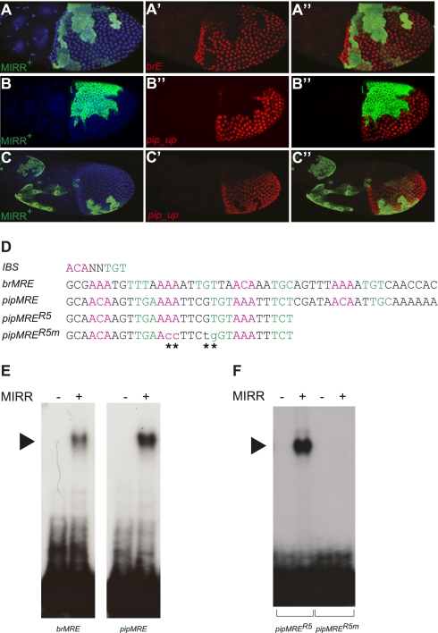
Epidermal growth factor receptor (EGFR) controls a wide range of developmental events, from body axes specification in insects to cardiac development in humans. During Drosophila oogenesis, a gradient of EGFR activation patterns the follicular epithelium. Multiple transcriptional targets of EGFR in this tissue have been identified, but their regulatory elements are essentially unknown. We report the regulatory elements of broad (br) and pipe (pip), two important targets of EGFR signaling in Drosophila oogenesis. br is expressed in a complex pattern that prefigures the formation of respiratory eggshell appendages. We found that this pattern is generated by dynamic activities of two regulatory elements, which display different responses to Pointed, Capicua, and Mirror, transcription factors involved in the EGFR-mediated gene expression. One of these elements is active in a pattern similar to pip, a gene repressed by EGFR and essential for establishing the dorsoventral polarity of the embryo. We demonstrate that this similarity of expression depends on a common sequence motif that binds Mirror in vitro and is essential for transcriptional repression in vivo.


Conflict of interest statement

The authors declare no conflict of interest.

Figures

Fig. 1.
Fig. 1.
br expression is regulated by two cis-regulatory modules. (A) Schematic of the genomic locus of br with genomic fragments used to generate transgenic reporter constructs depicted as bars. Gray bars indicate fragments with no enhancer activity during oogenesis and black bars denote fragments which activate patterned reporter gene expression (br4 and br6). Fragments brL and brE, used in all subsequent experiments, are shown in green and red, respectively. (B–E′′) BR protein expression compared with the expression of brE-lacZ and brL-EGFP reporters in egg chambers at stages 9, 10A, and 10B (lateral views, dorsal side up). Samples were stained with anti-BR antibody (magenta), anti–β-Gal antibody (red), anti-GFP antibody (green), and DAPI (blue) to visualize nuclei. Panels E–E′′ are merged images of brE-lacZ and brL-GFP reporter staining. (B–B′′) At stage 9, uniform BR expression (B) is mediated by brE (C), but the brL reporter is silent (D). (B–D′) At stage 10A, BR is cleared from the dorsal midline domain (B′). This pattern is formed by the combination of loss of brE-reporter expression in a wide dorsal domain (C′) and brL activating reporter expression in two distinct dorsolateral patches within the clearance of brE. (D′). At stage 10B, higher levels of BR are visible in the patches (B′′) produced by increasing activity of brL (D′′). (F–G′′) Reporter expression controlled by direct fusion of brL and brE fragments (brLE) fully recapitulates the dynamics of BR expression. Double immunostaining for brLE reporter expression (brLE-LacZ, anti–β-Gal antibody) and BR protein (αBR antibody). Nuclei stained by DAPI. Magnification, 20×. (H) Summary of the spatial and temporal contribution of the two CRMs to the dynamic changes of BR expression as derived from the profiling shown in B–G′′ and Fig. S1 (dorsal view). At any time point of egg shell development, expression of BR (magenta) is the sum of the expression activated by brE (red) and brL (green).
Fig. 2.
Fig. 2.
Effectors of EGFR signaling regulate brE and brL differentially. (A–F′′′ and G) Immunostaining for GFP (green, A–F), β-Gal (red, A–F′), and BR (gray, A′′′–F′′′) of stage 10B egg chambers carrying either brE-LacZ or brL-LacZ reporter (A–F). Mutant clones are marked by the loss of GFP. Yellow lines have been added to mark clone boundaries relevant for this analysis. G schematically summarizes the results of the mosaic experiments and illustrate the effects of clonal inactivation of pnt, mirr, and cic on the expression of brE, brL, and BR (clones represented within dotted back lines) (A–B′′′) pntΔ86 clones in the dorsal midline domain did not affect brE (29 of 30 clones), but produced ectopic expression of brL (10 of 11 clones). White arrowheads mark the position of the dorsal midline; dorsal views are shown. (C–D′′′) mirr1825 clones in dorsal follicle cells produced ectopic expression of brE (C and C′) (22 of 23 clones), and loss of brL expression in the appendage forming cells (D and D) (26 of 28 clones). (C′′ and D′′) GFP and β-Gal merged images show enlarged views of the anterior domain (clone areas 1 in both C and D). Dorsal views are shown. (E–F′′′) cicfetU6 clones in ventral anterior follicle cells produced loss of brE (109 of 115 clones; clone 2 in E), and ectopic expression of brL (11 of 11 clones; clone 2 in F), but dorsal anterior clones did not produce misexpression (clone 1 in E, and clone 1 in F). These effects were restricted to the anterior half of the egg chamber, as shown in clones spanning the anterior-posterior prepatterning boundary (clone 3 in E, and clone 2 in F; see also schematic in G). brL expression in cic clones was still suppressed in the anteriormost two to three rows of cells (arrowhead in F′′), potentially by the Dpp pathway. (E′′ and F′′) GFP and β-Gal merged images show enlarger views of the ventral anterior domain (lower left section in E and F, respectively). Lateral views with dorsal side up are shown; magnification, 20×. (H) Summary of the effects of EGFR signaling on the two CRMs of br. PNT acts solely on brL to repress its expression at dorsal most cells. MIRR, which becomes activated by EGFR signaling through the repression of CIC, represses brE and activates brL.
Fig. 3.
Fig. 3.
A short DNA sequence mediates dorsal repression of pip. (A) Genomic locus of pip with exons indicated in dark blue and intronic sequences in light blue pip_up and pip_down fragments used to generate reporter constructs are shown in red and gray, respectively. pipA (black bar) is the previously reported regulatory region of pip. The position of the identified MRE in pip_up is indicated by the arrowhead. (B) Sequence logo of the evolutionarily conserved motif present in both brE and pip_up. (C–D′) Reporter activity of stage 10 egg chambers (lateral view, dorsal up) from flies carrying either a pip_up-lacZ transgene (C) or a version of pip_up-lacZ lacking the 50-bp motif pip_upΔMRE (D) assessed by β-Gal immunostaining. Nuclei were stained by DAPI (C′ and D′); magnification, 20×. Although pip_up activates reporter expression in a pattern indistinguishable to that of endogenous pip, deletion of the motif results in ectopic expression of the reporter in dorsal follicle cells.
Fig. 4.
Fig. 4.
MIRR represses pip through direct binding to a MRE. (A–C) Lateral view of stage 10 egg chambers expressing MIRR in clones marked by coexpression of GFP (nuclei are shown in blue; magnification, 20×). Ectopic MIRR expression abolishes both brE expression (in 54 of 59 clones; A′) and pip_up expression (in 53 of 53 clones; B′ and C′, overlay in B′′ and C′′) in a cell-autonomous and position-independent manner. (D) Sequences of the Mirror response elements of br (brMRE) and pip (pipMRE). Nucleotide in green and magenta correspond to the two halves of the palindrome comprising the Iro binding site (IBS; ACANNTGT). pipMRER5 is a subfragment of pipMRE still able to bind to MIRR. pipMRER5m carries four nucleotide exchanges at positions that are conserved between the brMRE and the pipMRE (indicated by the asterisks). (E) EMSA with extracts expressing HA-tagged MIRR (+) or a control protein (−) and radioactively labeled brMRE or pipMRE probes. MIRR/DNA-complexes are indicated with the black arrowhead. (F) EMSA with control (−) or MIRR (+) expressing extracts and radioactively labeled pipMRER5 or pipMRER5m. Note that the four point mutations in pipMRER5m completely abolish MIRR binding.

Similar articles

Cited by

References

    1. Lynch JA, Peel AD, Drechsler A, Averof M, Roth S. EGF signaling and the origin of axial polarity among the insects. Curr Biol. 2010;20:1042–1047. - PMC - PubMed
    1. Moghal N, Sternberg PW. The epidermal growth factor system in Caenorhabditis elegans. Exp Cell Res. 2003;284:150–159. - PubMed
    1. Budi EH, Patterson LB, Parichy DM. Embryonic requirements for ErbB signaling in neural crest development and adult pigment pattern formation. Development. 2008;135:2603–2614. - PMC - PubMed
    1. Sanchez-Soria P, Camenisch TD. ErbB signaling in cardiac development and disease. Semin Cell Dev Biol. 2010;21:929–935. - PMC - PubMed
    1. Cheung LS, Schüpbach T, Shvartsman SY. Pattern formation by receptor tyrosine kinases: Analysis of the Gurken gradient in Drosophila oogenesis. Curr Opin Genet Dev. 2011;21:719–725. - PMC - PubMed

Publication types