. 2012 Jan 31;109(5):1578-83.
doi: 10.1073/pnas.1016741109. Epub 2012 Jan 17.

Homeobox gene distal-less is required for neuronal differentiation and neurite outgrowth in the Drosophila olfactory system

Affiliations

Homeobox gene distal-less is required for neuronal differentiation and neurite outgrowth in the Drosophila olfactory system

Jessica Plavicki et al. Proc Natl Acad Sci U S A. .

Abstract

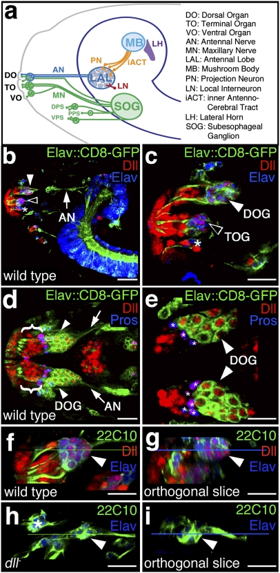
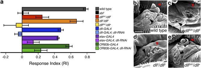
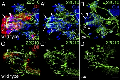
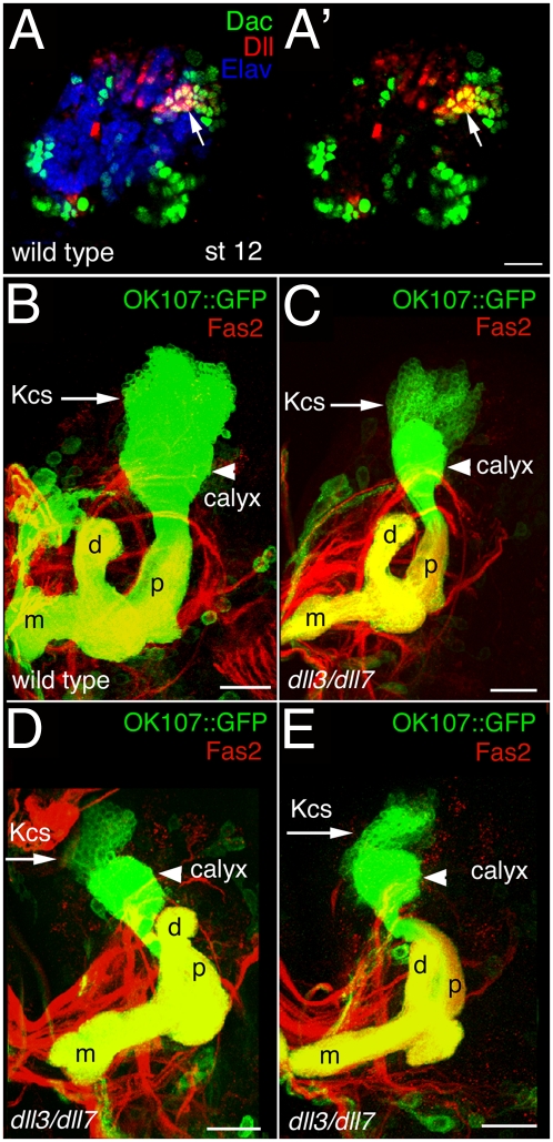
Vertebrate Dlx genes have been implicated in the differentiation of multiple neuronal subtypes, including cortical GABAergic interneurons, and mutations in Dlx genes have been linked to clinical conditions such as epilepsy and autism. Here we show that the single Drosophila Dlx homolog, distal-less, is required both to specify chemosensory neurons and to regulate the morphologies of their axons and dendrites. We establish that distal-less is necessary for development of the mushroom body, a brain region that processes olfactory information. These are important examples of distal-less function in an invertebrate nervous system and demonstrate that the Drosophila larval olfactory system is a powerful model in which to understand distal-less functions during neurogenesis.


Conflict of interest statement

The authors declare no conflict of interest.

Figures

Fig. 1.
Fig. 1.
dll is expressed in neurons and associated support cells during larval olfactory system development and is required for neuronal development. (A) Schematic of the larval chemosensory system (adapted from ref. 3). Neurons in the DOG, TOG, and VO ganglion (VOG) and the dorsal, posterior, and ventral pharyngeal sense organs (DPS, PPS, and VPS) respond to chemical compounds encountered in the environment and relay chemosensory information to target areas in the central nervous system. ORNs in the DOG (white arrowheads in BD) send afferent projections via the antennal nerve (AN; arrows in B and D) to the LAL where they form glomeruli connected by local interneurons (LNs). Projection neurons (PNs) of the LAL relay information via the inner antennocerebral tract (iACT) to brain areas associated with learning and memory: the lateral horn (LH) and MB. MN, maxillary nerve. (B and C) Lateral views at low (B) and high (C) magnification of a stage 16 elav-GAL4;UAS-mCD8-GFP Drosophila embryo stained for Dll (red) and the neuronal marker for embryonic lethal, abnormal vision (Elav; blue). GFP is localized to the membranes of neurons. Dll expression is detected in the DOG (white arrowheads in B and C), the TOG (open arrowheads in B and C), and the VOG (asterisk in C) as well as in support cells and epidermal cells. (D and E) Dorsal views at low (D) and high (E) magnification of a stage 16 elav-GAL4;UAS-mCD8-GFP Drosophila embryo stained for Dll (red) and Pros (blue). Dll is detected in DO neurons (white arrowheads in D and E) as well as the socket, sheath, and shaft support cells (brackets in D). Dll and Pros colocalize in sheath cells (purple; asterisks in E). (FI) Dorsal views of stage 16 30wild-type (F and G) and dll-null (H and I) embryos stained for sensory neurons (monoclonal antibody 22C10; green), Dll (red), and Elav (blue). The wild-type DOG (arrowheads in F and G) consists of ∼36 neurons (21 ORNs plus ∼15 GRNs), whereas dll-null embryos (arrowheads in H and I) have an average of 7 DOG neurons. Orthogonal slices through the corresponding confocal Z-series are shown in G and I. The horizontal green lines in F and H represent the planes of section for G and I. The horizontal blue lines in G and I represent the depths within each Z-series of the images shown F and H. The asterisk in H marks neurons that are not part of the DO. Absence of Dll staining was used to identify dll-null embryos. Anterior is to the left in all images. (BF and H) Optical sections (0.4 μm) were collected with a 40× objective. (C, E, F, and H) A 1.25× digital zoom was used. (Scale bars: 50 mm.)
Fig. 2.
Fig. 2.
dll mutants exhibit olfactory deficits in larval behavioral assays. (A) Olfactory RIs of dll mutants and dll-RNAi animals. See SI Materials and Methods for details of the assay. Data are plotted as mean ± SEM. With Student's two-tailed t test, P values <0.001 were obtained for dllSA1/dll3 (red histogram) and dllSA1/dll+ (yellow histogram) compared with wild type (gray histogram). Anosmic (sbl; black histogram) larvae served as a positive control. We also tested the responses of third-instar larvae in which UAS-dll-RNAi was driven by a pan-neural GAL4 driver (elav-GAL4; light purple histogram), an ORN-specific driver (OR83b-GAL4; light green histogram), or the dll-GAL4 driver (light blue histogram). Animals carrying any of the GAL4 drivers exhibited significant (P < 0.001) impairments in their olfactory behavioral responses even without the dll-RNAi construct present [dll-GAL4 (dark blue histogram), RI = 0.52 ± 0.08; elav-GAL4 (dark purple histogram), RI = 0.49 ± 0.02; and OR83b-GAL4 (dark green histogram), RI = 0.58 ± 0.04]. Nonetheless, when the dll-RNAi construct was present, the responses were significantly worse than with the respective GAL4 drivers alone (P < 0.05 for elav-GAL4 driving dll-RNAi and P < 0.001 for either OR83b-GAL4 or dll-GAL4 driving dll-RNAi). dll-GAL4 driving dll-RNAi (light blue histogram), RI = −0.05 ± 0.05; elav-GAL4 driving dll-RNAi (light purple histogram), RI = 0.15 ± 0.08; and OR83b-GAL4 driving dll-RNAi (light green histogram), RI = 0.25 ± 0.05. (BE) Scanning electron micrographs (1,200×) of third-instar larval DO (red arrowheads) and TO (black arrowheads) from wild-type (B), dllSA1/dll3 (C), dll1/dll3 (D), and dllSA1/dll+ (E) heads. dllSA1/dll3 larvae have malformations in the DO dome (C) and display significant impairments in behavioral assays (RI = 0.27 ± 0.03; red histogram in A). The DO dome is also malformed in dll1/dll3 larvae (C), but larvae still respond well to the attractive odorant (RI = 0.72 ± 0.09; orange histogram in A). In contrast, olfaction is significantly impaired in dllSA1/dll+ larvae (RI = −0.08 ± 0.16; yellow histogram in A) despite the proper formation of the cuticular components of the DO and TO (E). Anterior is at the top, and ventral midline is to the right. (Scale bar: 50 mm.)
Fig. 3.
Fig. 3.
Dendrite malformations, axon projection defects, and support cell loss in dll-null embryos. Lateral views of stage 16 wild-type (A and A') and dll-null (B) embryos stained for Cut (blue), Dll (red), and sensory neurons (monoclonal antibody 22C10; green). Cut is coexpressed with Dll in the majority of DOG support cells (white arrowheads), TOG support cells (open arrowheads), and VOG support cells and is weakly coexpressed in a few neurons in all three ganglia. In dll-null embryos, the number of support cells is decreased and the remaining support cells are disorganized (compare B with A and A'). Lateral views of stage 16 wild-type (C and C′) and dll-null (D) embryonic heads stained for neurons (monoclonal antibody 22C10; green) and Dll (red). Dendrites (white arrows) of the DOG (white arrowheads) are malformed in dll-null embryos. Dendrites (open arrows) of the TOG (open arrowheads) are also abnormal. The axons from both olfactory and GRNs of the DOG initially project via the antennal nerve (white tracing in C). However, before reaching the brain, ORN and GRN axons normally defasciculate, with ORNs innervating the LAL and GRNs projecting to the SOG. Axons from the TOG and the VOG form the maxillary nerve (blue tracing in C), which also normally targets the SOG. In dll-null animals, afferent projections from the DOG and TOG form a single fascicle directed toward the SOG (asterisks in B and D). Absence of Dll staining was used to identify dll-null embryos in B and D. (AB) Three-dimensional reconstructions of optical sections (0.4 μm) were collected with a 40× objective and a 1.4× digital zoom. (CD) Optical sections (0.4 μm) were collected with a 40× objective. Anterior is to the left in all images. (Scale bars: 50 mm.)
Fig. 4.
Fig. 4.
Loss of dll function disrupts the MBs. (A and A′) Lateral view of an embryonic stage 12 brain stained for Dac (green), Dll (red), and Elav (blue). Arrows point to young Kenyon cells, a subset of which coexpress Dll and Dac. (BE) Frontal views of late third-instar MBs of wild-type (B) and dll3/dll7 (CE) larvae carrying OK107-GAL4 driving UAS-mCD8-GFP (green) and stained with Fas2 antibody (red). The midline is toward the left. Arrows indicate the Kenyon cell clusters (Kcs), which are greatly reduced in the dll mutants. Arrowheads indicate the calyces, which also are reduced in the dll mutants. d, dorsal lobe; m, medial lobe; p, peduncle. The lobes and peduncles are thinner in the dll mutants, and the relative position of the peduncle and dorsal lobe is abnormal in the dll mutants. (Scale bars: 50 mm.)

Similar articles

Cited by

References

    1. Panganiban G, Rubenstein JL. Developmental functions of the Distal-less/Dlx homeobox genes. Development. 2002;129:4371–4386. - PubMed
    1. Panganiban G, et al. The origin and evolution of animal appendages. Proc Natl Acad Sci USA. 1997;94:5162–5166. - PMC - PubMed
    1. Vosshall LB, Stocker RF. Molecular architecture of smell and taste in Drosophila. Annu Rev Neurosci. 2007;30:505–533. - PubMed
    1. Gerber B, Stocker RF. The Drosophila larva as a model for studying chemosensation and chemosensory learning: A review. Chem Senses. 2007;32(1):65–89. - PubMed
    1. Singh R, Singh K. Fine structure of the sensory organs of Drosophila melanogaster Meigen larva (Diptera: Drosophilidae) Int J Insect Morphol Embryol. 1984;13(4):255–273.

Publication types

LinkOut - more resources