. 2012 Jan 31;109(5):1619-24.
doi: 10.1073/pnas.1108464109. Epub 2012 Jan 17.

Toll-like receptor 5 (TLR5), IL-1β secretion, and asparagine endopeptidase are critical factors for alveolar macrophage phagocytosis and bacterial killing

Affiliations

Toll-like receptor 5 (TLR5), IL-1β secretion, and asparagine endopeptidase are critical factors for alveolar macrophage phagocytosis and bacterial killing

Delphyne Descamps et al. Proc Natl Acad Sci U S A. .

Abstract

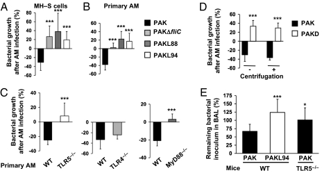
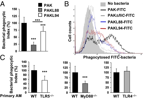
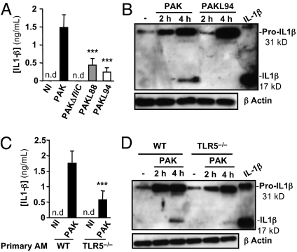
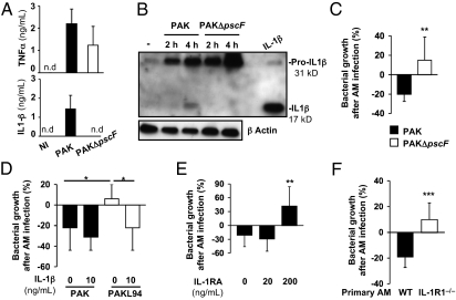
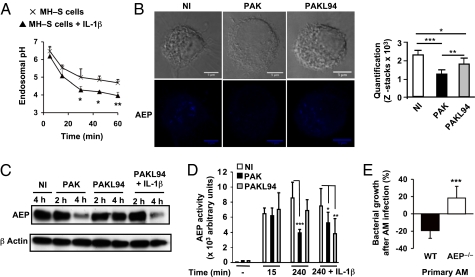
A deficit in early clearance of Pseudomonas aeruginosa (P. aeruginosa) is crucial in nosocomial pneumonia and in chronic lung infections. Few studies have addressed the role of Toll-like receptors (TLRs), which are early pathogen associated molecular pattern receptors, in pathogen uptake and clearance by alveolar macrophages (AMs). Here, we report that TLR5 engagement is crucial for bacterial clearance by AMs in vitro and in vivo because unflagellated P. aeruginosa or different mutants defective in TLR5 activation were resistant to AM phagocytosis and killing. In addition, the clearance of PAK (a wild-type P. aeruginosa strain) by primary AMs was causally associated with increased IL-1β release, which was dramatically reduced with PAK mutants or in WT PAK-infected primary TLR5(-/-) AMs, demonstrating the dependence of IL-1β production on TLR5. We showed that this IL-1β production was important in endosomal pH acidification and in inducing the killing of bacteria by AMs through asparagine endopeptidase (AEP), a key endosomal cysteine protease. In agreement, AMs from IL-1R1(-/-) and AEP(-/-) mice were unable to kill P. aeruginosa. Altogether, these findings demonstrate that TLR5 engagement plays a major role in P. aeruginosa internalization and in triggering IL-1β formation.


Conflict of interest statement

The authors declare no conflict of interest.

Figures

Fig. 1.
Fig. 1.
P. aeruginosa clearance by AMs is dependent on efficient TLR5-Flagella interaction. MH–S cells (A) or primary AMs (B) were infected for 4 h with WT PAK, PAKΔfliC, PAKL88, or PAKL94 mutants [MOI: 0.1 (B) or 10 (A)]. (C) Primary WT, TLR5−/−, TLR4−/−, or MyD88−/− AMs were infected for 4 h with WT PAK (MOI: 0.1). (D) Primary AMs were infected for 4 h with either WT PAK or PAKD (MOI: 0.1) with or without centrifugation. (AD) CFU were quantified in AM supernatants pooled with cell lysates. Results are means ± SD of three experiments (**P < 0.01; ***P < 0.001) and are expressed as percentages as follows: (CFU counts recovered without AMs − CFU counts recovered after AM infection) × 100. (E) WT or TLR5−/− mice were infected intratracheally with 105 CFU of PAK or PAKL94. BALs were performed 2 h later and plated on LB agar plates to quantify total CFU counts. Results are means ± SD of three experiments (*P < 0.05; ***P < 0.001) and are expressed as percentage of surviving bacteria in BAL = (total CFU counts in BAL/ CFU counts of inoculum) × 100.
Fig. 2.
Fig. 2.
Phagocytosis of P. aeruginosa by AMs is dependent on TLR5-MyD88 signaling. (A and C) MH–S cells were infected for 1 h with WT PAK, PAKL88, or PAKL94 mutants (A), or primary WT, TLR5−/−, MyD88−/−, or TLR4−/− AMs were infected with WT PAK (MOI: 10) for 1 h (C). After stimulation, cells were washed and treated with tobramycin and the number of ingested bacteria was determined by counting CFU from AM lysates on LB agar plates. Results are means ± SD of three experiments (***P < 0.001) and are expressed as percentages of relative phagocytosis index = (CFU counts in mutant PAK-treated cells/CFU counts in WT PAK-treated cells) × 100. (B) FACS analysis of MH–S cells following 1 h infection with FITC-labeled WT or FITC-mutant P. aeruginosa strains. Results are representative of three independent experiments.
Fig. 3.
Fig. 3.
IL-1β production in primary AMs is dependent on TLR5. A total of 105 primary AMs from WT (A and B) or TLR5−/− (C and D) mice were infected with WT PAK, PAKL88, or PAKL94 (MOI: 1). IL-1β secretion was measured in supernatants 4 h postinfection (A and C). Results are means ± SD of three experiments (***P < 0.001). NI, noninfected cells; nd, not detected. Lysates obtained from primary WT (B) or TLR5−/− (D) AMs infected with bacteria were analyzed by immunoblotting for pro-IL-1β processing. Equal loading was controlled for by β-actin detection. Results are representative of three independent experiments.
Fig. 4.
Fig. 4.
IL-1β secretion is required for AMs to kill P. aeruginosa. (A) Primary WT AMs (105) were infected with WT PAK or PAKΔpscF mutant (MOI: 1) for 4 h. TNFα or IL-1β was measured in supernatants. Results are means ± SD of three experiments. NI, noninfected cells; nd, not detected. (B) Lysates from infected AMs were assessed for pro-IL-1β processing. Equal loading was controlled by β-actin detection. Results are representative of three independent experiments. (C) Primary WT AMs were infected with WT PAK or PAKΔpscF (MOI: 0.1) for 4 h. (D and E) MH–S cells were infected with WT PAK or PAKL94 mutant (MOI: 0.1) for 4 h in the presence or not of IL-1β or IL-1 receptor antagonist (IL-1RA). (F) Primary WT or IL-1R1−/− AMs were infected for 4 h with WT PAK (MOI: 0.1). (CF) CFU were quantified in supernatants pooled with cell lysates. Results are means ± SD of three experiments (*P < 0.05; **P < 0.01) and are expressed as in Fig 1.
Fig. 5.
Fig. 5.
AEP protease is essential for P. aeruginosa killing by primary AMs. (A) Kinetics of endolysosomal pH in MH–S cells ± IL-1β (10 ng/mL) was measured. Results are means ± SD of three experiments. (B) Confocal microscopy of AEP was measured in primary WT AMs infected or not for 3 h with WT PAK or PAKL94. Quantification was performed using Image J software (n = 5). (C) Immunoblotting AEP expression in lysates from WT PAK- or PAKL94-infected primary WT AMs (MOI: 1) with or without IL-1β. Equal loading was controlled by β-actin detection. Results are representative of three independent experiments. (D) AEP activity was measured in lysates from WT PAK- or PAKL94-infected MH–S cells (MOI: 10) with a fluorometer by assessing the hydrolysis of the specific substrate for AEP. (AD) Unpaired t test (*P < 0.01; **P < 0.005; ***P < 0.0001). NI, noninfected cells. (E) Primary WT or AEP−/− AMs were infected with WT PAK (MOI: 0.1) for 4 h. CFUs were quantified in supernatants pooled with cell lysates. Results are means ± SD of three experiments (***P < 0.001) and are expressed as in Fig 1.

Similar articles

Cited by

References

    1. Mogayzel PJ, Jr, Flume PA. Update in cystic fibrosis 2009. Am J Respir Crit Care Med. 2010;181:539–544. - PubMed
    1. Gordon S. The macrophage: Past, present and future. Eur J Immunol. 2007;37(Suppl 1):S9–S17. - PubMed
    1. Di A, et al. CFTR regulates phagosome acidification in macrophages and alters bactericidal activity. Nat Cell Biol. 2006;8:933–944. - PubMed
    1. Deriy LV, et al. Disease-causing mutations in the cystic fibrosis transmembrane conductance regulator determine the functional responses of alveolar macrophages. J Biol Chem. 2009;284:35926–35938. - PMC - PubMed
    1. Kawai T, Akira S. The role of pattern-recognition receptors in innate immunity: Update on Toll-like receptors. Nat Immunol. 2010;11:373–384. - PubMed

Publication types

LinkOut - more resources