. 2012 Jan 31;109(5):1637-42.
doi: 10.1073/pnas.1120790109. Epub 2012 Jan 17.

Cooperative effects of aminopeptidase N (CD13) expressed by nonmalignant and cancer cells within the tumor microenvironment

Affiliations

Cooperative effects of aminopeptidase N (CD13) expressed by nonmalignant and cancer cells within the tumor microenvironment

Liliana Guzman-Rojas et al. Proc Natl Acad Sci U S A. .

Abstract

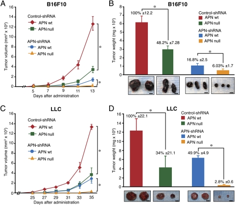
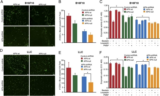
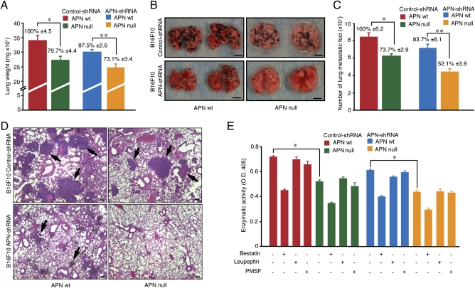
Processes that promote cancer progression such as angiogenesis require a functional interplay between malignant and nonmalignant cells in the tumor microenvironment. The metalloprotease aminopeptidase N (APN; CD13) is often overexpressed in tumor cells and has been implicated in angiogenesis and cancer progression. Our previous studies of APN-null mice revealed impaired neoangiogenesis in model systems without cancer cells and suggested the hypothesis that APN expressed by nonmalignant cells might promote tumor growth. We tested this hypothesis by comparing the effects of APN deficiency in allografted malignant (tumor) and nonmalignant (host) cells on tumor growth and metastasis in APN-null mice. In two independent tumor graft models, APN activity in both the tumors and the host cells cooperate to promote tumor vascularization and growth. Loss of APN expression by the host and/or the malignant cells also impaired lung metastasis in experimental mouse models. Thus, cooperation in APN expression by both cancer cells and nonmalignant stromal cells within the tumor microenvironment promotes angiogenesis, tumor growth, and metastasis.


Conflict of interest statement

The authors declare no conflict of interest.

Figures

Fig. 1.
Fig. 1.
Loss of APN expression by tumor or host nonmalignant cells impairs tumor growth. Control and APN shRNA B16F10 or LLC cells were injected into the right flank of WT or APN-null mice (n = 5/group), and tumor growth was followed. (A and C) Average tumor volume (mm3) was measured every 2 d (*P < 0.02). (B and D) Tumor weights were evaluated after 13 d (B16F10) or 35 d (LLC). Bars represent mean tumor weight ± SEM (*P < 0.02). Photographs show images of representative tumors. (Scale bar, 5 mm.)
Fig. 2.
Fig. 2.
Reduction of blood vessel density in tumors correlates with reduction of APN activity. (A and D) Immunofluorescence analysis of sections from B16F10 or LLC tumors (as indicated) collected at the study end point was performed with anti-CD31 and secondary FITC-conjugated antibodies (green). (Scale bar, 100 μm.) (B and E) Quantification of blood vessel numbers by manual counting (16 randomly chosen 10× fields per group) in APN WT and APN knockout (null) mice injected with APN knockdown (shRNA) or control-shRNA. (C and F) Enzymatic activity of APN in protein extracts derived from APN WT and APN-null mice carrying control and APN-shRNA B16F10 tumors. Soluble tumor protein extracts were incubated with l-leucine-p-nitroanilide substrate in the presence (+) or absence (−) of enzymatic inhibitors (bestatin, leupeptin, and PMSF). The bars represent mean values of enzymatic activity ± SEM from triplicates (*P < 0.03).
Fig. 3.
Fig. 3.
Disruption of APN expression in tumor and/or nonmalignant stromal cells affects formation of lung metastases. B16F10 melanoma cells (control and APN-shRNA) were injected into a tail vein of WT and APN-null mice. Lungs (n = 5) were dissected 3 wk later. (A) Lung weights are shown as means ± SEM (*P < 0.01; **P < 0.006). (B) Representative photographs of lungs. (Scale bar, 5 mm.) (C) Metastatic foci were counted on lungs of WT and APN-null mice (*P < 0.01; **P < 0.002). (D) Formalin-fixed lung sections were stained with H&E. Black arrows indicate lung metastatic foci. (Scale bar, 100 μm.) (E) Enzymatic activity of APN was evaluated in lungs with metastasis. Bars represent means ± SEM (*P < 0.0001).

References

    1. Dixon J, et al. Expression of aminopeptidase-n (CD 13) in normal tissues and malignant neoplasms of epithelial and lymphoid origin. J Clin Pathol. 1994;47:43–47. - PMC - PubMed
    1. Stange T, Kettmann U, Holzhausen HJ. Immunoelectron microscopic single and double labelling of aminopeptidase N (CD 13) and dipeptidyl peptidase IV (CD 26) Acta Histochem. 1996;98:323–331. - PMC - PubMed
    1. Mina-Osorio P. The moonlighting enzyme CD13: Old and new functions to target. Trends Mol Med. 2009;9:449–456. - PMC - PubMed
    1. Favaloro EJ, Moraitis N, Bradstock K, Koutts J. Co-expression of haemopoietic antigens on vascular endothelial cells: A detailed phenotypic analysis. Br J Haematol. 1990;74:385–394. - PubMed
    1. Ashmun RA, Look AT. Metalloprotease activity of CD13/aminopeptidase N on the surface of human myeloid cells. Blood. 1990;75:462–469. - PubMed

Publication types