. 2012 Jan 31;109(5):1643-8.
doi: 10.1073/pnas.1120985109. Epub 2012 Jan 17.

Oncogene-specific activation of tyrosine kinase networks during prostate cancer progression

Affiliations

Oncogene-specific activation of tyrosine kinase networks during prostate cancer progression

Justin M Drake et al. Proc Natl Acad Sci U S A. .

Abstract

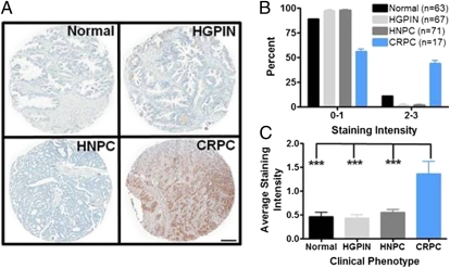
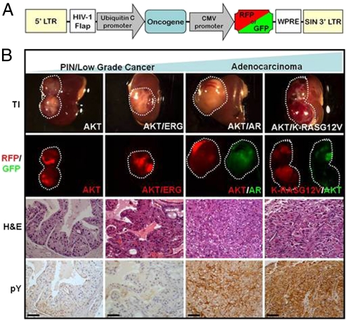
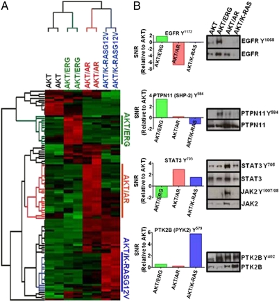
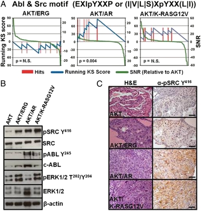
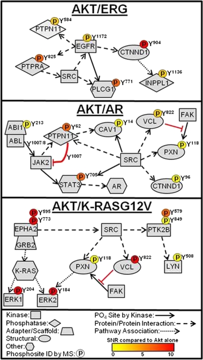
Dominant mutations or DNA amplification of tyrosine kinases are rare among the oncogenic alterations implicated in prostate cancer. We demonstrate that castration-resistant prostate cancer (CRPC) in men exhibits increased tyrosine phosphorylation, raising the question of whether enhanced tyrosine kinase activity is observed in prostate cancer in the absence of specific tyrosine kinase mutation or DNA amplification. We generated a mouse model of prostate cancer progression using commonly perturbed non-tyrosine kinase oncogenes and pathways and detected a significant up-regulation of tyrosine phosphorylation at the carcinoma stage. Phosphotyrosine peptide enrichment and quantitative mass spectrometry identified oncogene-specific tyrosine kinase signatures, including activation of EGFR, ephrin type-A receptor 2 (EPHA2), and JAK2. Kinase:substrate relationship analysis of the phosphopeptides also revealed ABL1 and SRC tyrosine kinase activation. The observation of elevated tyrosine kinase signaling in advanced prostate cancer and identification of specific tyrosine kinase pathways from genetically defined tumor models point to unique therapeutic approaches using tyrosine kinase inhibitors for advanced prostate cancer.


Conflict of interest statement

The authors declare no conflict of interest.

Figures

Fig. 1.
Fig. 1.
Robust phosphotyrosine expression is observed in castration-resistant prostate cancer (CRPC) specimens. (A) Representative image of immunohistochemical staining using the phosphotyrosine-specific antibody, 4G10, of prostate specimens ranging from normal to CRPC. Tissue spots from patients with CRPC show high levels of phosphotyrosine expression in the epithelial compartment. (B) Increased tyrosine phosphorylation is observed in CRPC, with nearly 50% of the patients displaying high-intensity staining (2, 3) compared with normal, HGPIN, or HNPC tissues. (C) Average staining intensity of all of the tissues clearly show a significant increase of tyrosine phosphorylation in CRPC patients. HGPIN, high-grade prostatic intraepithelial neoplasia; HNPC, hormone naïve prostate cancer; HRPC, hormone refractory prostate cancer. ***P < 0.001, one-way ANOVA. (Scale bar, 200 μm.)
Fig. 2.
Fig. 2.
Phosphotyrosine expression is increased during prostate cancer progression. (A) Lentiviral vector diagram displaying the organization of oncogene and fluorescent marker expression used in these tumors. (B) Gross and histological morphology of each tumor type after 12-wk engraftment in SCID mice using the prostate regeneration protocol. Fluorescence corresponds to expression of a particular oncogene. IHC staining of progressive mouse tumor phenotypes reveals an increasing gradient of phosphotyrosine expression with more aggressive tumors expressing higher levels than indolent tumors. TI, transillumination; H&E, hematoxylin and eosin; pY, phosphotyrosine. (Scale bars, 50 μm.)
Fig. 3.
Fig. 3.
Unique phosphotyrosine signatures are observed in a mouse model of prostate cancer progression. (A) Heatmap representing unique clusters of tyrosine phosphorylation for each mouse tumor phenotype. Each row corresponds to a unique phosphopeptide. Red, hyperphosphorylation; green, hypophosphorylation for each phosphopeptide. (B) Specific tyrosine kinases are observed in an oncogene-specific fashion. Signal-to-noise ratio (SNR) (relative to AKT) was calculated for each phosphorylation event and plotted. Positive SNR confirms elevation of that particular phosphorylation event. Western blotting validates indicated oncogene-specific phosphorylation results.
Fig. 4.
Fig. 4.
Bioinformatic analysis reveals enrichment of dasatinib tyrosine kinase targets in AKT/AR tumors. (A) Enrichment analysis of tyrosine phosphosite motifs reveals enrichment of phosphosubstrates of the tyrosine kinases ABL and SRC, targets of the tyrosine kinase inhibitor dasatinib, in AKT/AR tumors. No significant enrichment of these phosphopeptides were observed in either AKT/ERG or AKT/K-RASG12V tumors. Enrichment scores for all kinase motifs are shown in Dataset S2. (B) Western blot and (C) IHC staining for the activated kinases ABL, SRC, or ERK1/2 reveal tumor-specific activation of these kinases. (Scale bars, 50 μm.)
Fig. 5.
Fig. 5.
Curation of phosphoproteomic profiling and bioinformatics delineates distinct tyrosine kinase signaling pathways in an oncogene-specific manner. Selected substrate and interaction pathways from each tyrosine kinase were generated from a combination of our phosphoproteomics dataset and the HPRD and Phosphosite databases. An elevated phosphorylation event identified by MS is indicated by a phosphorylation residue depicted above the protein and color coded. Solid arrow, protein is a direct substrate of the upstream kinase at that site. Dashed arrow, protein interacts directly with the upstream kinase/protein. Dotted arrow, protein is found within the pathway of the upstream kinase/protein.

Similar articles

Cited by

References

    1. Nakahara M, et al. A novel gain-of-function mutation of c-kit gene in gastrointestinal stromal tumors. Gastroenterology. 1998;115:1090–1095. - PubMed
    1. Ben-Neriah Y, Daley GQ, Mes-Masson AM, Witte ON, Baltimore D. The chronic myelogenous leukemia-specific P210 protein is the product of the bcr/abl hybrid gene. Science. 1986;233:212–214. - PubMed
    1. Dienstmann R, Martinez P, Felip E. Personalizing therapy with targeted agents in non-small cell lung cancer. Oncotarget. 2011;2:165–177. - PMC - PubMed
    1. Kim KS, et al. Predictors of the response to gefitinib in refractory non-small cell lung cancer. Clin Cancer Res. 2005;11:2244–2251. - PubMed
    1. Druker BJ, et al. Activity of a specific inhibitor of the BCR-ABL tyrosine kinase in the blast crisis of chronic myeloid leukemia and acute lymphoblastic leukemia with the Philadelphia chromosome. N Engl J Med. 2001;344:1038–1042. - PubMed

Publication types