. 2012 Jan 31;109(5):1673-8.
doi: 10.1073/pnas.1115884109. Epub 2012 Jan 17.

IL-33 is processed into mature bioactive forms by neutrophil elastase and cathepsin G

Affiliations

IL-33 is processed into mature bioactive forms by neutrophil elastase and cathepsin G

Emma Lefrançais et al. Proc Natl Acad Sci U S A. .

Abstract

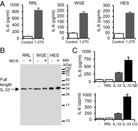
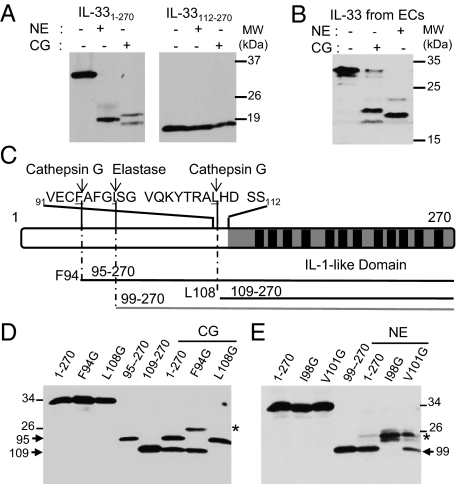
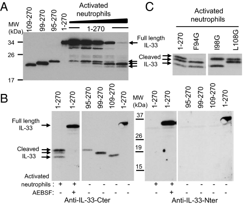
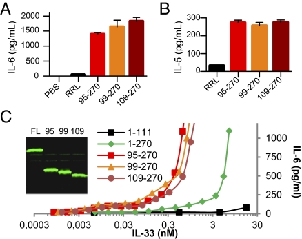
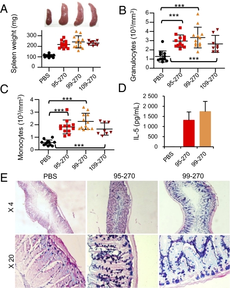
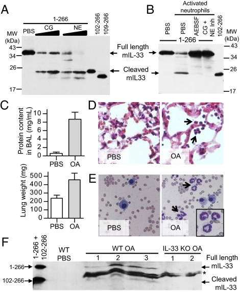
Interleukin-33 (IL-33) (NF-HEV) is a chromatin-associated nuclear cytokine from the IL-1 family, which has been linked to important diseases, including asthma, rheumatoid arthritis, ulcerative colitis, and cardiovascular diseases. IL-33 signals through the ST2 receptor and drives cytokine production in type 2 innate lymphoid cells (ILCs) (natural helper cells, nuocytes), T-helper (Th)2 lymphocytes, mast cells, basophils, eosinophils, invariant natural killer T (iNKT), and natural killer (NK) cells. We and others recently reported that, unlike IL-1β and IL-18, full-length IL-33 is biologically active independently of caspase-1 cleavage and that processing by caspases results in IL-33 inactivation. We suggested that IL-33, which is released upon cellular damage, may function as an endogenous danger signal or alarmin, similar to IL-1α or high-mobility group box 1 protein (HMGB1). Here, we investigated the possibility that IL-33 activity may be regulated by proteases released during inflammation. Using a combination of in vitro and in vivo approaches, we demonstrate that neutrophil serine proteases cathepsin G and elastase can cleave full-length human IL-33(1-270) and generate mature forms IL-33(95-270), IL-33(99-270), and IL-33(109-270). These forms are produced by activated human neutrophils ex vivo, are biologically active in vivo, and have a ~10-fold higher activity than full-length IL-33 in cellular assays. Murine IL-33 is also cleaved by neutrophil cathepsin G and elastase, and both full-length and cleaved endogenous IL-33 could be detected in the bronchoalveolar lavage fluid in an in vivo model of acute lung injury associated with neutrophil infiltration. We propose that the inflammatory microenvironment may exacerbate disease-associated functions of IL-33 through the generation of highly active mature forms.


Conflict of interest statement

The authors declare no conflict of interest.

Figures

Fig. 1.
Fig. 1.
The biological activity of full-length IL-331–270 is increased by neutrophil elastase and cathepsin G. (A) Capacity of full-length IL-331–270 produced in RRL, WGE, or HES (5 μL of lysate) to activate the IL-33-responsive mast cell line MC/9 (2 × 10⁵ cells/well) was analyzed by determining IL-6 levels in supernatants using an ELISA. (B) Western blot analysis (305B mAb) of full-length IL-331–270 produced in RRL, WGE, or HES before or after incubation with MC/9 cells for 24 h. (C) Full-length IL-331–270 produced in RRL (5 μL of lysate) was preincubated with neutrophil elastase (NE) (30 mU/μL; 30 min at 37 °C) or cathepsin G (CG) (0.09 mU/μL; 1 h at 37 °C) before incubation with MC/9 cells (105 cells/well) for 24 h. IL-6 levels in supernatants were detected by ELISA. Results in A and C are shown as means and SD of three separate data points and are representative of two independent experiments.
Fig. 2.
Fig. 2.
Neutrophil elastase and cathepsin G generate IL-33 mature forms IL-3395–270, IL-3399–270, and IL-33109–270. (A and B) IL-33 is a substrate for neutrophil elastase and cathepsin G. In vitro-translated IL-331–270 and IL-33112–270 proteins (A) or endogenous native IL-33 isolated from necrotic endothelial cells (B) were incubated with purified neutrophil elastase (NE) (30 mU/μL; 30 min at 37 °C) or cathepsin G (CG) (0.09 mU/μL; 1 h at 37 °C). (C) Primary structure of human IL-33. The IL-1-like domain with its 12 β-strands (black boxes) is indicated. The sequence surrounding the cathepsin G and elastase cleavage sites is shown. (D and E) Identification of the cathepsin G and elastase cleavage sites using IL-33 single-point and deletion mutants. Mutation of F94 and L108 to glycine abrogates formation of the 21- and 18-kDa CG (0.06 mU/μL; 1 h at 37 °C) cleavage products, respectively (D). Mutation of I98 to glycine abrogates formation of the 20 kDa elastase (19 mU/μL; 30 min at 37 °C) cleavage product (E). In vitro-translated proteins IL-3395–270, IL-33109–270, and IL-3399–270 comigrate on SDS/PAGE with the 21- and 18-kDa cathepsin G cleavage products (D) and the 20-kDa elastase cleavage product (E), respectively. *, secondary cleavage products. Proteins were separated by SDS/PAGE and revealed by Western blot with anti-IL-33-Cter mAb 305B (AE). Blots are representative of at least three independent experiments.
Fig. 3.
Fig. 3.
Activated human neutrophils generate IL-33 mature forms IL-3395–270, IL-3399–270, and IL-33109–270. (A and B) In vitro-translated full-length human IL-331–270 was incubated with PMA-activated human neutrophils. Proteins were comigrated on SDS/PAGE with in vitro-translated IL-3395–270, IL-3399–270, and IL-33109–270 proteins and revealed by Western blot with anti-IL-33-Cter mAb 305B (A and B, Left) or anti-IL-33-Nter antibodies (B, Right). IL-33 was incubated with increasing amounts of supernatants from activated neutrophils (6 × 10⁴–106 neutrophils; 15 min at 37 °C) (A). Cleavage of IL-33 by activated neutrophils (105 neutrophils; 2 h at 37 °C) was inhibited by serine protease inhibitor AEBSF (1 mM) (B). (C) In vitro-translated IL-331–270 or single-point mutants IL-33F94G, IL-33I98G, and IL-33L108G were incubated with PMA-activated human neutrophils (105 neutrophils; 2 h at 37 °C). Proteins were separated on SDS/PAGE and revealed by Western blot with anti-IL-33-Cter mAb 305B. Blots are representative of three independent experiments, with neutrophils isolated from three different donors.
Fig. 4.
Fig. 4.
IL-33 mature forms IL-3395–270, IL-3399–270, and IL-33109–270 are biologically active. (A and B) The capacity of IL-33 mature forms produced in RRL (5 μL of lysate) to activate the IL-33-responsive MC/9 mast cells (105 cells/well; 24-h stimulation) (A) and KU812 basophil-like chronic myelogenous leukemia cells (5 × 10⁵ cells/well; 24-h stimulation) (B) was analyzed by determining cytokine levels in supernatants [IL-6 (A); IL-5 (B)] using ELISAs. Results are shown as means and SD of three separate data points. (C) Comparison of the biological activity of IL-33 full-length and mature forms. The different proteins were quantified by fluorescence and various concentrations were used to stimulate MC/9 mast cells (105 cells/well; 24-h stimulation). IL-6 protein levels in supernatants were detected by ELISA. Results are representative of at least two independent experiments.
Fig. 5.
Fig. 5.
In vivo activity of IL-33 mature forms IL-3395–270, IL-3399–270, and IL-33109–270. (AE) BALB/c mice were injected (i.p.) with recombinant proteins (4 μg) every day during 1 wk, and spleens (A), peripheral blood (B and C), serum (D), and jejunum (E) were collected at day 8 (n = 12/3 experiments for PBS, IL-3395–270, and IL-3399–270; n = 8/2 experiments for IL-33109–270). Spleen weights and representative photographs for each group of mice are shown (A). Numbers of peripheral blood granulocytes (B), monocytes (C), and serum concentrations of IL-5 (D) were determined. ***P < 0.001 (Student unpaired and two-tailed t test). Epithelial changes in the jejunum are shown (E). Mucus was stained with periodic acid Schiff and alcian blue.
Fig. 6.
Fig. 6.
Murine IL-33 is processed by neutrophil elastase and cathepsin G ex vivo, and cleaved endogenous IL-33 is detected in vivo in a model of acute lung injury associated with neutrophil infiltration. (A and B) In vitro-translated full-length murine IL-331–266 (2.5 μL) was incubated with purified neutrophil cathepsin G (CG) (14.7 and 29.4 μU/μL) or elastase (NE) (7.3 and 14.7 mU/μL) (A) or with supernatants of activated mouse bone marrow neutrophils (3 × 10⁴ neutrophils) (B) for 1 h at 37 °C. Activated neutrophils were pretreated with PBS, serine protease inhibitor AEBSF (8 mM), or cathepsin G and elastase inhibitors (50 μM) for 1 h at 37 °C. Proteins were comigrated on SDS/PAGE with in vitro-translated murine IL-33102–266 and IL-33109–266 and revealed by Western blot with goat anti-mouse IL-33-Cter antibodies. Blots are representative of at least three independent experiments. (CF) Full-length and cleaved endogenous IL-33 are released in BAL fluid following OA-induced acute lung injury. Lung injury was assessed by measuring lung weight and protein content in BAL fluid two hours after i.v. OA administration (C). Accumulation of neutrophils (black arrows) was analyzed by hematoxylin/eosin staining of lung tissue sections (D) and May–Grunwald–Giemsa staining of BAL fluids cytospins (E). Proteins from cell-free BAL fluids (30 μL) of OA-treated wild-type (three mice) or IL-33−/− mice (two mice) were comigrated on SDS/PAGE with in vitro-translated murine IL-331–266 and IL-33102–266 and revealed by Western blot with goat anti-mouse IL-33-Cter antibodies (F). *Nonspecific bands detected in both wild-type and IL-33−/− mice.

Similar articles

Cited by

References

    1. Dinarello CA. Immunological and inflammatory functions of the interleukin-1 family. Annu Rev Immunol. 2009;27:519–550. - PubMed
    1. Sims JE, Smith DE. The IL-1 family: regulators of immunity. Nat Rev Immunol. 2010;10:89–102. - PubMed
    1. Dinarello CA. Interleukin-1 in the pathogenesis and treatment of inflammatory diseases. Blood. 2011;117:3720–3732. - PMC - PubMed
    1. Schmitz J, et al. IL-33, an interleukin-1-like cytokine that signals via the IL-1 receptor-related protein ST2 and induces T helper type 2-associated cytokines. Immunity. 2005;23:479–490. - PubMed
    1. Baekkevold ES, et al. Molecular characterization of NF-HEV, a nuclear factor preferentially expressed in human high endothelial venules. Am J Pathol. 2003;163:69–79. - PMC - PubMed

Publication types