. 2012 Jan 31;109(5):1679-84.
doi: 10.1073/pnas.1117885109. Epub 2012 Jan 17.

Syndecan-4 proteoliposomes enhance fibroblast growth factor-2 (FGF-2)-induced proliferation, migration, and neovascularization of ischemic muscle

Affiliations

Syndecan-4 proteoliposomes enhance fibroblast growth factor-2 (FGF-2)-induced proliferation, migration, and neovascularization of ischemic muscle

Eugene Jang et al. Proc Natl Acad Sci U S A. .

Abstract

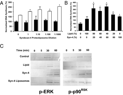
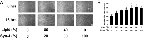
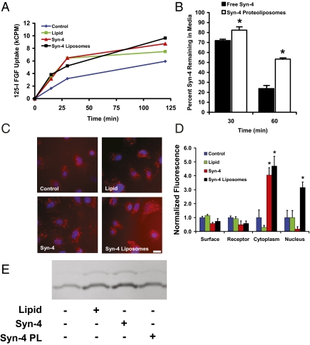
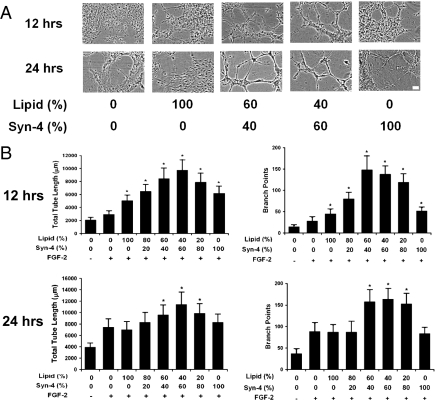
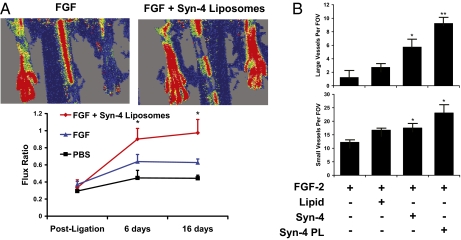
Ischemia of the myocardium and lower limbs is a common consequence of arterial disease and a major source of morbidity and mortality in modernized countries. Inducing neovascularization for the treatment of ischemia is an appealing therapeutic strategy for patients for whom traditional treatment modalities cannot be performed or are ineffective. In the past, the stimulation of blood vessel growth was pursued using direct delivery of growth factors, angiogenic gene therapy, or cellular therapy. Although therapeutic angiogenesis holds great promise for treating patients with ischemia, current methods have not found success in clinical trials. Fibroblast growth factor-2 (FGF-2) was one of the first growth factors to be tested for use in therapeutic angiogenesis. Here, we present a method for improving the biological activity of FGF-2 by codelivering the growth factor with a liposomally embedded coreceptor, syndecan-4. This technique was shown to increase FGF-2 cellular signaling, uptake, and nuclear localization in comparison with FGF-2 alone. Delivery of syndecan-4 proteoliposomes also increased endothelial proliferation, migration, and angiogenic tube formation in response to FGF-2. Using an animal model of limb ischemia, syndecan-4 proteoliposomes markedly improved the neovascularization following femoral artery ligation and recovery of perfusion of the ischemic limb. Taken together, these results support liposomal delivery of syndecan-4 as an effective means to improving the potential of using growth factors to achieve therapeutic neovascularization of ischemic tissue.


Conflict of interest statement

Conflict of interest statement: The authors declare a conflict of interest. A.B.B. and E.R.E. have filed a patent on the technology discussed in this paper.

Figures

Fig. 1.
Fig. 1.
Syndecan-4 proteoliposomes enhance FGF-2–induced proliferation, migration, and intracellular signaling. (A) Syndecan-4 proteoliposomes induce enhanced endothelial proliferation in response to FGF-2 treatment. Black bars are samples without FGF-2 and white bars are samples treated with 10 ng/mL FGF-2. (B) Midrange composition (lipid-to-protein ratio) proteoliposomes were optimally active in inducing enhanced endothelial proliferation. (C) Western blotting analysis of cells treated with 10 ng/mL FGF-2 shows increased and prolonged phosphorylation of MAPK with syndecan-4 or syndecan-4 proteoliposome treatment. Phosphorylation of p90RSK was increased in endothelial cells treated with FGF-2 in combination with syndecan-4 or syndecan-4 proteoliposomes *Statistically different from FGF-2 alone treatment group (P < 0.05). All Western blots were performed in duplicate from separate experiments with similar results.
Fig. 2.
Fig. 2.
In vitro migration and wound closure is increased by FGF-2 treatment in combination with syndecan-4 proteoliposomes. Confluent monolayers of endothelial cells were wounded by scraping with a plastic cell scraper. Wound closure and cell migration was examined at various times with phase-contrast microscopy. (A) Quantitative analysis of wound edge closure following wounding and in the presence of various treatments. (B) Wound-healing assay shows increased wound edge migration with syndecan-4 proteoliposome treatment. (Scale bar, 100 μm.) *Statistically significant difference between samples (P < 0.05).
Fig. 3.
Fig. 3.
Syndecan-4 proteoliposomes enhance FGF-2 uptake and intracellular trafficking. (A) Liposomally embedded syndecan-4 increases uptake of 125I-labeled FGF-2 in endothelial cells. FGF-2 alone (blue line), FGF-2 with liposomes (green line), FGF-2 with syndecan-4 (red line), and FGF-2 with syndecan-4 proteoliposomes (black line). (B) Liposome embedding prolongs 3H-labeled syndecan-4 presence in the media after 30 or 60 min. Syndecan-4 alone is shown with black bars, and syndecan-4 proteoliposomes are shown with white bars. (C) Uptake of fluorescently labeled FGF-2 is increased with syndecan-4 proteoliposomes. FGF-2 was labeled with Texas Red dye and added to cultures of endothelial cells for 4 h. (Scale bar, 20 μm.) (D) Nuclear localization of FGF-2 is increased in the presence of syndecan-4 proteoliposomes. Endothelial cells were exposed to fluorescently labeled FGF-2 for 4 h. The cells were then fractionated and the fluorescence in each fraction was measured and corrected for autofluorescence. (E) Lipids, syndecan-4, and syndecan-4 proteoliposomes enhance rac-1 activity. Cells were treated with various compounds and subjected to a pull-down assay for the active GTP-bound form of Rac-1.
Fig. 4.
Fig. 4.
Syndecan-4 proteoliposomes enhance in vitro tube formation in combination with FGF-2. HUVECs were seeded on extracellular matrix and exposed to various treatments. In vitro tube formation was quantified by measuring total tube length and the number of branch points. All samples were treated with FGF-2 (10 ng/mL) unless labeled otherwise. (A) Phase-contrast micrographs of endothelial cells in Matrigel after 12 h of treatment. (Scale bar, 20 μm.) (B) Quantitative analysis of tube length formed by endothelial network after 12 h of treatment. Number of branch points in endothelial networks after 12 h. Length of tubes in the endothelial network following 24 h of incubation. Branch points in endothelial networks after 24 h of treatment. *Statistically different from FGF-2 group (P < 0.05).
Fig. 5.
Fig. 5.
Enhanced neovascularization of ischemic muscle after treatment with syndecan-4 proteoliposomes in combination with FGF-2. Hind-limb ischemia was created in rats through femoral artery ligation, and treatments were delivered over 7 d with an osmotic pump. (A) Routine histological staining revealed reduced ischemic changes following treatment with FGF-2 in combination with syndecan-4 alone or with liposomally embedded syndecan-4. (Scale bar, 100 μm.) (B) Quantification of large vessel number per field of view. Quantification of capillary number per field of view. *Statistically different from FGF group (P < 0.05). **Statistically different from all other groups (P < 0.05).

Similar articles

Cited by

References

    1. Tirziu D, Simons M. Angiogenesis in the human heart: Gene and cell therapy. Angiogenesis. 2005;8:241–251. - PubMed
    1. Simons M, et al. Pharmacological treatment of coronary artery disease with recombinant fibroblast growth factor-2: Double-blind, randomized, controlled clinical trial. Circulation. 2002;105:788–793. - PubMed
    1. Bikfalvi A, Klein S, Pintucci G, Rifkin DB. Biological roles of fibroblast growth factor-2. Endocr Rev. 1997;18:26–45. - PubMed
    1. Nugent MA, Iozzo RV. Fibroblast growth factor-2. Int J Biochem Cell Biol. 2000;32:115–120. - PubMed
    1. Rapraeger AC, Krufka A, Olwin BB. Requirement of heparan sulfate for bFGF-mediated fibroblast growth and myoblast differentiation. Science. 1991;252:1705–1708. - PubMed

Publication types

LinkOut - more resources