. 2012 Jan 31;109(5):1697-701.
doi: 10.1073/pnas.1114476109. Epub 2012 Jan 17.

Polar growth in the Alphaproteobacterial order Rhizobiales

Affiliations

Polar growth in the Alphaproteobacterial order Rhizobiales

Pamela J B Brown et al. Proc Natl Acad Sci U S A. .

Erratum in

  • Proc Natl Acad Sci U S A. 2012 Feb 21;109(8);3190

Abstract

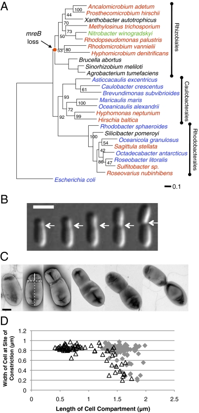
Elongation of many rod-shaped bacteria occurs by peptidoglycan synthesis at discrete foci along the sidewall of the cells. However, within the Rhizobiales, there are many budding bacteria, in which new cell growth is constrained to a specific region. The phylogeny of the Rhizobiales indicates that this mode of zonal growth may be ancestral. We demonstrate that the rod-shaped bacterium Agrobacterium tumefaciens grows unidirectionally from the new pole generated after cell division and has an atypical peptidoglycan composition. Polar growth occurs under all conditions tested, including when cells are attached to a plant root and under conditions that induce virulence. Finally, we show that polar growth also occurs in the closely related bacteria Sinorhizobium meliloti, Brucella abortus, and Ochrobactrum anthropi. We find that unipolar growth is an ancestral and conserved trait among the Rhizobiales, which includes important mutualists and pathogens of plants and animals.


Conflict of interest statement

The authors declare no conflict of interest.

Figures

Fig. 1.
Fig. 1.
Phylogeny and morphology of A. tumefaciens are suggestive of polar growth. (A) Maximum likelihood phylogeny inferred from gyrA sequences. Taxon label colors indicate bacteria described as reproducing by budding (red), binary fission (blue), budding or binary fission (green), and bacteria for which the mode of reproduction is not clearly described (black) (40). Node values indicate clade frequency among 100 nonparametric bootstrapped datasets. The scale bar indicates the estimated number of substitutions per site. The node where mreB (and the associated mreCD, rodA, pbpB genes) is predicted to have been lost within the Rhizobiales is indicated. See SI Materials and Methods for details of phylogenetic reconstruction. (B) Time-lapse microscopy of a single A. tumefaciens cell reveals that constriction of A. tumefaciens cells occurs before cell division. White arrows indicate the site of constriction. Images shown were taken at 30-min intervals. (Scale bar: 2 μm.) (C) Transmission electron micrographs of individual A. tumefaciens cells at different stages in the cell cycle. The lines in the second panel illustrate metrics used in quantification. Solid line is length of daughter cell compartment, dashed line is the width of the cell at the site of constriction, and dotted line is the length of the mother cell compartment. (Scale bar: 0.5 μm.) (D) Width of cells at the site of constriction is plotted against the length of the daughter cell compartment (open black triangles) and length of the mother cell compartment (gray diamonds).
Fig. 2.
Fig. 2.
FtsZ-eGFP localizes to the early constriction in A. tumefaciens. (A) FtsZ-eGFP localization in wild type A. tumefaciens. (B) Time-lapse microscopy of a single A. tumefaciens cell expressing FtsZ-eGFP during growth. (C) FtsZ-eGFP localization in TRSE-stained A. tumefaciens cells after a short (15-min) chase. (D) FtsZ-eGFP localization in TRSE-stained A. tumefaciens cells after a longer (2-h) chase.
Fig. 3.
Fig. 3.
A. tumefaciens cells display zonal cell wall synthesis. (A) Time-lapse microscopy of growth of a single E. coli cell pulse labeled with TRSE. (B) Growth pattern of a young cell pulse labeled with TRSE and d-cys labeling patterns of individual cell sacculi. Illustrations of the d-cys-labeled sacculi are provided. (C) Growth pattern of a predivisional cell pulse labeled with TRSE and d-cys-labeling patterns of individual cell sacculi. Illustrations of the d-cys-labeled sacculi are provided. (White scale bars: 2 μm; black scale bars: 0.5 μm.)
Fig. 4.
Fig. 4.
Model for the segregation of peptidoglycan and outer membrane labels in A. tumefaciens cells. The illustration shows the expected patterns for the distribution of labeled peptidoglycan or outer membrane in cells of different ages at the initiation of the chase, as indicated above each panel. Red indicates the area of the cell retaining the original label. Yellow indicate areas of active PG synthesis. Areas of new, unlabeled PG are shown in orange. The location of the label will remain constant in newly generated daughter cells due to the conservative nature of the growth process. The labeling and subsequent growth of intermediate and predivisional cells clearly explains generation of cells (and sacculi) with well-defined thin or thick bands of labeled material, respectively.
Fig. 5.
Fig. 5.
Unipolar growth is conserved among the Rhizobiales. (A) d-cys labeling of individual S. meliloti sacculi. (Scale bar: 0.5 μm.) (B) Time-lapse microscopy of TRSE-stained Brucella abortus 544 cells. (C) Time-lapse microscopy of TRSE-stained O. anthropi cells. (D) TRSE-stained H. denitrificans cells after 18 h. (E) Time-lapse microscopy of TRSE-stained P. hirschii cells. (White scale bars: 2 μm.)

References

    1. den Blaauwen T, de Pedro MA, Nguyen-Distèche M, Ayala JA. Morphogenesis of rod-shaped sacculi. FEMS Microbiol Rev. 2008;32:321–344. - PubMed
    1. de Pedro MA, Quintela JC, Höltje JV, Schwarz H. Murein segregation in Escherichia coli. J Bacteriol. 1997;179:2823–2834. - PMC - PubMed
    1. Chauhan A, et al. Interference of Mycobacterium tuberculosis cell division by Rv2719c, a cell wall hydrolase. Mol Microbiol. 2006;62:132–147. - PubMed
    1. Flärdh K. Essential role of DivIVA in polar growth and morphogenesis in Streptomyces coelicolor A3(2) Mol Microbiol. 2003;49:1523–1536. - PubMed
    1. Kang CM, Nyayapathy S, Lee JY, Suh JW, Husson RN. Wag31, a homologue of the cell division protein DivIVA, regulates growth, morphology and polar cell wall synthesis in mycobacteria. Microbiology. 2008;154:725–735. - PubMed

Publication types

MeSH terms