Comparative Study
. 2012 Apr 1;302(7):H1355-66.
doi: 10.1152/ajpheart.00918.2011. Epub 2012 Feb 3.

Vena cava and aortic smooth muscle cells express transglutaminases 1 and 4 in addition to transglutaminase 2

Affiliations
Comparative Study

Vena cava and aortic smooth muscle cells express transglutaminases 1 and 4 in addition to transglutaminase 2

Kyle B Johnson et al. Am J Physiol Heart Circ Physiol. .

Abstract

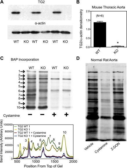
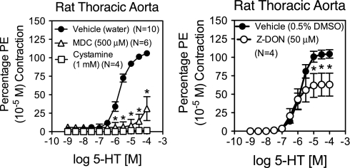
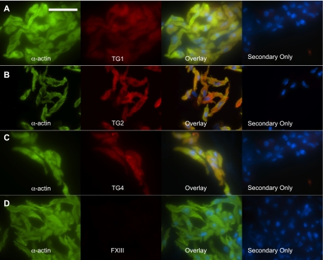
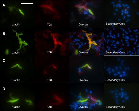
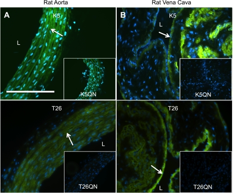
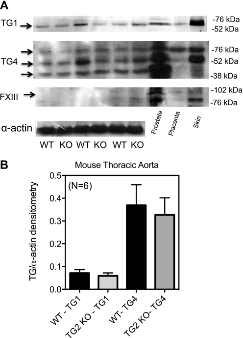
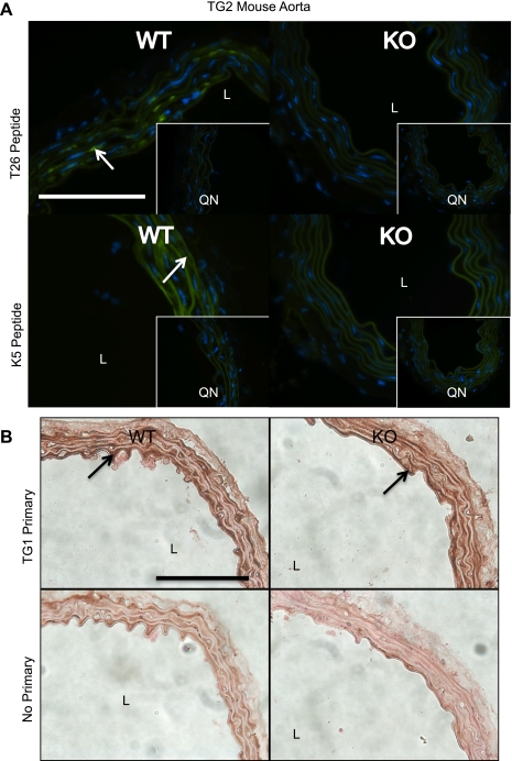
Transglutaminase (TG) function facilitates several vascular processes and diseases. Although many of these TG-dependent vascular processes have been ascribed to the function of TG2, TG2 knockout mice have a mild vascular phenotype. We hypothesized that TGs besides TG2 exist and function in the vasculature. Biotin-pentylamide incorporation, a measure of general TG activity, was similar in wild-type and TG2 knockout mouse aortae, and the general TG inhibitor cystamine reduced biotin-pentylamine incorporation to a greater extent than the TG2-specific inhibitor Z-DON, indicating the presence of other functional TGs. Additionally, 5-hydroxytryptamine-induced aortic contraction, a TG-activity-dependent process, was decreased to a greater extent by general TG inhibitors vs. Z-DON (maximum contraction: cystamine = abolished, monodansylcadaverine = 28.6 ± 14.9%, and Z-DON = 60.2 ± 15.2% vehicle), providing evidence for the importance of TG2-independent activity in the vasculature. TG1, TG2, TG4, and Factor XIII (FXIII) mRNA in rat aortae and vena cavae was detected by RT-PCR. Western analysis detected TG1 and TG4, but not FXIII, in rat aortae and vena cavae and in TG2 knockout and wild-type mouse aortae. Immunostaining confirmed the presence of TG1, TG2, and TG4 in rat aortae and vena cavae, notably in smooth muscle cells; FXIII was absent. K5 and T26, FITC-labeled peptide substrates specific for active TG1 and TG2, respectively, were incorporated into rat aortae and vena cavae and wild-type, but not TG2 knockout, mouse aortae. These studies demonstrate that TG2-independent TG activity exists in the vasculature and that TG1 and TG4 are expressed in vascular tissues.


Figures

Fig. 1.
Fig. 1.
Transglutaminase (TG) activity in aortic tissues that lack TG2 or in the presence of TG inhibitors. A: Western analysis of TG2 protein shows that TG2 is essentially abolished in the TG2 knockout (KO) mouse vs. wild-type (WT) aorta. B: densitometry of TG2 in WT and KO mouse aortae. Bars represent the means ± SE of 6 different WT and KO mice. *P < 0.05. C: biotin-pentylamide (BAP) incorporation, a measure of TG activity, shows that the TG2 KO mouse still has significant levels of TG activity that are similar to those of WT. Blot represents aortae from 4 different TG2 WT and KO mouse pairs. Bottom: analysis of the blot in C using Un-Scan-It to determine the relative intensity of each BAP blot. Peaks represent different bands detected in the blot. Numbers next to the bands on the blots corresponds to the peak of the same number in the graph to the left. Green lines represent a WT animal and yellow a KO animal. Red lines represent the same WT animal in the presence of 1 mmol/l cystamine and blue lines a KO animal in the presence of cystamine. D: BAP incorporation into proteins of normal rat aorta homogenates in the presence of vehicle, 1 mmol/l cystamine, or 50 μmol/l Z-DON. BAP incorporation was reduced by both cystamine and Z-DON; however, BAP incorporation in the presence of cystamine was notably less than in the presence of Z-DON.
Fig. 2.
Fig. 2.
Rat aorta contraction to 5-hydroxytryptamine (5-HT) in the presence of TG inhibitors. The ability of rat aortae to contract to 5-HT was tested in the presence of TG inhibitors or vehicle. All inhibitors were able to reduce contraction of the aorta compared with vehicle. Global TG inhibitor cystamine (1 mmol/l) abolished contraction to 5-HT, while monodansylcadavarine (MDC; 500 μmol/l) significantly reduced contraction (maximum contraction = 28.6 ± 14.9% of vehicle). The TG2-specific inhibitor Z-DON (50 μmol/l) only slightly reduced contraction to 5-HT (maximum contraction = 60.2 ± 15.2% of vehicle). Contraction is reported as a percentage of initial contraction to phenylephrine (PE). *P ≤ 0.05, significantly reduced from vehicle.
Fig. 3.
Fig. 3.
Western analysis of normal rat aorta and vena cava tissues. A: immunoblotting with anti-TG antibodies detects TG1, TG2, and TG4 in aorta and vena cava tissue homogenates but not Factor XIII (FXIII). +, Positive control (rat skin lysate for TG1, human prostate lysate for TG4 and HeLa cell lysate for FXIII). Each lane represents an aorta or vena cava homogenate derived from a different animal for a total of 4 animals. B: densitometry of TG1, TG2, and TG4 protein found in blots in A. Normalization of the TGs to β-actin indicates significantly more TG1 and TG2, but not TG4, is present in vena cava compared with aorta tissues (means ± SE: aorta TG1 = 0.51 ± 0.05, TG2 = 0.80 ± 0.02, TG4 = 0.18 ± 0.01; vena cava TG1 = 0.842 ± 0.069, TG2 = 1.1 ± 0.1, TG4 = 0.48 ± 0.08). Arrows point to bands used for quantification. *P ≤ 0.05, significantly different from vena cava.
Fig. 4.
Fig. 4.
Immunohistochemical staining of TGs in normal rat aorta and vena cava. A: TG1 in rat aorta and vena cava tissues. Human skin was used as a positive control. B: TG4 in rat aorta and vena cava tissues. Human prostate was used as a positive control. C: FXIII in rat aorta and vena cava tissues. Human placenta was used as a positive control. Representative of tissues from four different animals. Arrows point to regions of staining. No primary denotes that primary antibody was left out of the reaction. L, lumen of the vessel. Scale bar = 100 μm.
Fig. 5.
Fig. 5.
TGs in freshly dissociated rat aorta smooth muscle cells. Rat thoracic aorta tissues were enzymatically dissociated to yield vascular smooth muscle cells. Confirmation that cells were smooth muscle cells was made by staining the cells with FITC-conjugated smooth muscle cell specific α-actin (green). Immunocytochemical detection of the TGs (red) demonstrated the presence of TG1 (A), TG2 (B), and TG4 (C) but not FXIII (D). Overlaying the red and green channels shows colocalization (yellow) of TG1, TG2, and TG4 with α-actin. Secondary only denotes cells from the same tissue dissociation that were incubated with secondary antibody but not primary antibodies. Representative of tissue dissociations from 4 different animals. Scale bar = 50 μm. Nuclei were stained with DAPI (blue).
Fig. 6.
Fig. 6.
TGs in freshly dissociated rat vena cava smooth muscle cells. Rat vena cava tissues were enzymatically dissociated to yield vascular smooth muscle cells. Confirmation that cells were smooth muscle cells was made by staining the cells with FITC-conjugated smooth muscle cell specific α-actin (green). Immunocytochemical detection of the TGs (red) demonstrated the presence of TG1 (A), TG2 (B), and TG4 (C) in smooth muscle cells. D: FXIII (red) was occasionally detected in the dissociation but was absent in smooth muscle cells (green). Overlaying the red and green channels shows colocalization (yellow) of TG1, TG2, and TG4 with α-actin. Secondary only denotes cells from the same tissue dissociation that were incubated with secondary antibody but not primary antibodies. Representative of tissue dissociations from 4 different animals. Scale bar = 50 μm. Nuclei are stained with DAPI (blue).
Fig. 7.
Fig. 7.
In situ detection of TG activity in rat aorta and vena cava. Sections of fresh frozen rat aorta (A) and vena cava (B) were tested for their ability to incorporate FITC-labeled peptides that are specific for TG1 (0.1 μmol/l K5) or TG2 (1 μmol/l T26) activity, demonstrating the presence of active TGs in these tissues. Mutant peptides (0.1 μmol/l K5QN and 1 μmol/l T26QN) that consist of the same amino acids, except that the active glutamine has been replaced with an asparagine and thus does not act as a substrate for the active enzymes, were used as negative controls. Representative of tissue sections from 3 different animals. Arrows point to areas of TG activity. Scale bar = 100 μm. Nuclei are stained with DAPI (blue).
Fig. 8.
Fig. 8.
TG isoform expression in the aorta of WT and TG2 KO mice. A: Western analysis of TG1, TG4, and FXIII in the TG2 KO and WT mouse. B: densitometric analysis of TG1 and TG4 in the TG2 KO mouse shows no statistical difference in the level of protein present between these 2 animals. Level of TG protein was compared with the amount of α-actin present. Arrows point to bands used for densitometry. Data represent aortae taken from 6 different WT and KO TG2 mice. P ≤ 0.05.
Fig. 9.
Fig. 9.
In situ detection of TG activity and immunohistochemical detection of TG1 in WT and TG2 KO mouse aortae. A: sections of fresh frozen sections of aorta from WT and TG2 KO mice were assayed for their ability to incorporate K5 (TG1 activity) or T26 (TG2 activity). Representative of aortic sections from 5 WT and 5 KO mice. K5QN and T26QN are the mutant, negative control peptides for TG1 and TG2, respectively. Arrow points to areas of TG activity. Nuclei are stained with DAPI. B: immunohistochemical analysis shows that TG1 is present in aorta from both the WT (top) and TG2 KO (bottom) mice. Arrows point to darkened areas of TG1 staining. Representative of tissues from 5 different WT and TG2 KO mice. Scale bars = 100 μm. Nuclei are stained with DAPI (blue).

Similar articles

Cited by

References

    1. Bailey CD, Johnson GV. The protective effects of cystamine in the R6/2 Huntington's disease mouse involve mechanisms other than the inhibition of tissue transglutaminase. Neurobiol Aging 27: 871–879, 2006. - PubMed
    1. Bakker EN, Buus CL, Spaan JA, Perree J, Ganga A, Rolf TM, Sorop O, Bramsen LH, Mulvany MJ, Vanbavel E. Small artery remodeling depends on tissue-type transglutaminase. Circ Res 96: 119–126, 2005. - PubMed
    1. Bakker EN, Pistea A, Spaan JA, Rolf T, de Vries CJ, van Rooijen N, Candi E, VanBavel E. Flow-dependent remodeling of small arteries in mice deficient for tissue-type transglutaminase: possible compensation by macrophage-derived factor XIII. Circ Res 99: 86–92, 2006. - PubMed
    1. Bakker EN, Pistea A, VanBavel E. Transglutaminases in vascular biology: relevance for vascular remodeling and atherosclerosis. J Vasc Res 45: 271–278, 2008. - PubMed
    1. Baumgartner W, Golenhofen N, Weth A, Hiiragi T, Saint R, Griffin M, Drenckhahn D. Role of transglutaminase 1 in stabilisation of intercellular junctions of the vascular endothelium. Histochem Cell Biol 122: 17–25, 2004. - PubMed

Publication types

MeSH terms