. 2013 Jan;12(1):5-17.
doi: 10.1007/s10237-012-0377-8. Epub 2012 Feb 4.

Calcific nodule morphogenesis by heart valve interstitial cells is strain dependent

Affiliations

Calcific nodule morphogenesis by heart valve interstitial cells is strain dependent

Charles I Fisher et al. Biomech Model Mechanobiol. 2013 Jan.

Abstract

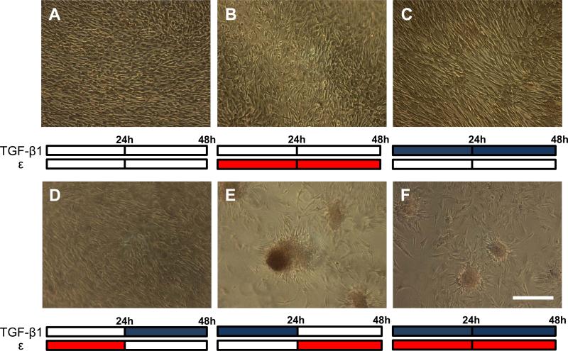
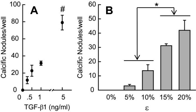
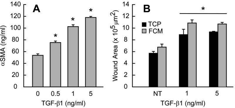
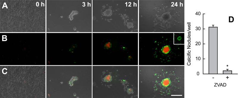
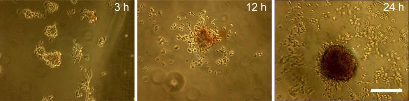
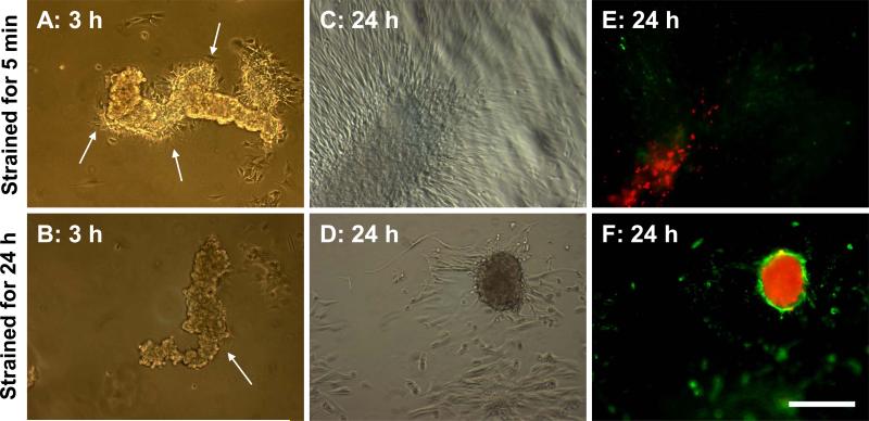
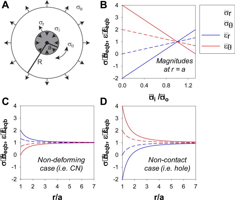
Calcific aortic valve disease (CAVD) results in impaired function through the inability of valves to fully open and close, but the causes of this pathology are unknown. Stiffening of the aorta is associated with CAVD and results in exposing the aortic valves to greater mechanical strain. Transforming growth factor β1 (TGF-β1) is enriched in diseased valves and has been shown to combine with strain to synergistically alter aortic valve interstitial cell (AVIC) phenotypes. Therefore, we investigated the role of strain and TGF-β1 on the calcification of AVICs. Following TGF-β1 pretreatment, strain induced intact monolayers to aggregate and calcify. Using a wound assay, we confirmed that TGF-β1 increases tension in the monolayer in parallel with α-smooth muscle actin (αSMA) expression. Continual exposure to strain accelerates aggregates to calcify into mature nodules that contain a necrotic core surrounded by an apoptotic ring. This phenotype appears to be mediated by strain inhibition of AVIC migration after the initial formation of aggregates. To better interpret the extent to which externally applied strain physically impacts this process, we modified the classical Lamé solution, derived using principles from linear elasticity, to reveal strain magnification as a novel feature occurring in a mechanical environment that supports nodule formation. These results indicate that strain can impact multiple points of nodule formation: by modifying tension in the monolayer, remodeling cell contacts, migration, apoptosis, and mineralization. Therefore, strain-induced nodule formation provides new directions for developing strategies to address CAVD.


Figures

Fig. 1
Fig. 1
Calcific nodule formation following combination treatments of 15% equibiaxial strain (red bars) and 1 ng/ml of TGF-β1 (blue bars) over 48 hrs. Control (A), strain only (B), and TGF-β1 only (C) all show unperturbed monolayers, as does strain followed by TGF-β1 (D). TGF-β1 followed by strain reveals large, mature calcific nodules (E), while simultaneous TGF-β1 and strain cause small, less mature nodules (F). All treatments stained with Alizarin red to identify calcification. This experiment was independently replicated three times with identical results. Scale bar = 250 μm.
Fig. 2
Fig. 2
Nodule formation as a function of TGF-β1 concentration (A) and strain magnitude (B). For A, 24 h of 15% equibiaxial strain was applied after 24 h of each TGF-β1 concentration. For B, all groups were treated with 1 ng/ml TGF-β1 for 24 h prior to each strain being applied for 24 h. All bar graphs and points represent mean ± SE (n≥3), * = p<0.05, # = p<0.01.
Fig. 3
Fig. 3
(A) aSMA expression increases with increasing dose of TGF-β1. (B) Mechanical damage reveals TGF-β1 effects on monolayer integrity. Monolayers were scraped with a 200 μl pipette tip in a cross pattern. Initial wound area increases with TGF-β1 pretreatment on tissue culture plastic (TCP) and Flexcell membrane (FCM). All bar graphs and points represent mean ± SE (n≥2), * = p<0.05.
Fig. 4
Fig. 4
Nodule maturation spreads from the inside out and is dependent on apoptosis. Cultures were removed from strain at 3, 12, and 24 h and stained with Annexin V and propidium iodide (red) to identify apoptosis and necrosis respectively. Phase only (A), fluorescence only (B), phase and fluorescence overlay (C). Lack of stain at 0 h indicates cell viability and monolayer integrity. Stain intensity increases with time as apoptosis is replaced by an intense necrotic core. Inset represents green channel only showing apoptotic ring at the nodule periphery. Inhibition of apoptosis with ZVAD reduces the number of mature nodules per well (D). This experiment was independently replicated three times with identical results. * = p<0.05. Scale bar = 250 μm.
Fig. 5
Fig. 5
Nodules calcification occurs concomitantly with maturation. Cultures were removed from strain at 3, 12, and 24 h and stained with alizarin red. Stain increases intensity with time. No fixation was used prior to staining causing cells to round up. A minimum of 3 wells were observed for each case. Scale bar = 250 μm.
Fig. 6
Fig. 6
Nodule maturation is strain dependent. Aggregation was induced with exposure to 5 min of strain. Cultures were either removed from strain (A, C, E) or continued to receive cyclic strain for 24 h (B, D, F). Images were collected at 3 and 24 h and stained with Annexin V and propidium iodide (red) to identify apoptosis and necrosis respectively. Arrows point to AVIC migration emerging from the aggregate but seem to be inhibited with strain. Flattened aggregates with minimal apoptotic staining (E) were not observed in cultures under 24 h strain. This experiment was independently replicated three times with identical results. Scale bar = 250 μm.
Fig. 7
Fig. 7
Strain enhancement model: Modified Lamé solution. External (σo) and internal (σi) stresses applied to a ring (radius = R) surrounding a circular core (radius = a) (A) used to calculate radial and circumferential stresses and strains at the core periphery (r = a) normalized to the equibiaxial case (B). Normalized gradient of stresses and strains as a function of the core radius were calculated for the non-deforming case (σi = 4σo/3) (C) and the non-contact case (σi = 0) (D). For all cases, R >> a. Normalized magnitudes indicate the extent of magnification of applied stresses and strains at the periphery of the circular core suggesting how small increases in strain can have significant impact on cells.

References

    1. Balachandran K, Konduri S, Sucosky P, Jo H, Yoganathan AP. An ex vivo study of the biological properties of porcine aortic valves in response to circumferential cyclic stretch. Ann Biomed Eng. 2006;34(11):1655–1665. doi:10.1007/s10439-006-9167-8. - PMC - PubMed
    1. Balachandran K, Sucosky P, Jo H, Yoganathan AP. Elevated cyclic stretch alters matrix remodeling in aortic valve cusps: implications for degenerative aortic valve disease. Am J Physiol Heart Circ Physiol. 2009;296(3):H756–764. doi:00900.2008 [pii] 10.1152/ajpheart.00900.2008. - PubMed
    1. Balachandran K, Sucosky P, Jo H, Yoganathan AP. Elevated cyclic stretch induces aortic valve calcification in a bone morphogenic protein-dependent manner. Am J Pathol. 2010;177(1):49–57. doi:ajpath.2010.090631 [pii] 10.2353/ajpath.2010.090631. - PMC - PubMed
    1. Balestrini JL, Skorinko JK, Hera A, Gaudette GR, Billiar KL. Applying controlled non-uniform deformation for in vitro studies of cell mechanobiology. Biomech Model Mechanobiol. 2010;9(3):329–344. doi:10.1007/s10237-009-0179-9. - PubMed
    1. Benton JA, Kern HB, Anseth KS. Substrate properties influence calcification in valvular interstitial cell culture. J Heart Valve Dis. 2008;17(6):689–699. - PMC - PubMed

Publication types

Substances

Supplementary concepts

LinkOut - more resources