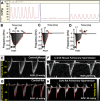
A brief overview of mouse models of pulmonary arterial hypertension: problems and prospects
- PMID: 22307907
- PMCID: PMC3774477
- DOI: 10.1152/ajplung.00362.2011
A brief overview of mouse models of pulmonary arterial hypertension: problems and prospects
Abstract
Many chronic pulmonary diseases are associated with pulmonary hypertension (PH) and pulmonary vascular remodeling, which is a term that continues to be used to describe a wide spectrum of vascular abnormalities. Pulmonary vascular structural changes frequently increase pulmonary vascular resistance, causing PH and right heart failure. Although rat models had been standard models of PH research, in more recent years the availability of genetically engineered mice has made this species attractive for many investigators. Here we review a large amount of data derived from experimental PH reports published since 1996. These studies using wild-type and genetically designed mice illustrate the challenges and opportunities provided by these models. Hemodynamic measurements are difficult to obtain in mice, and right heart failure has not been investigated in mice. Anatomical, cellular, and genetic differences distinguish mice and rats, and pharmacogenomics may explain the degree of PH and the particular mode of pulmonary vascular adaptation and also the response of the right ventricle.
Figures


Similar articles
-
Swine model of chronic postcapillary pulmonary hypertension with right ventricular remodeling: long-term characterization by cardiac catheterization, magnetic resonance, and pathology.J Cardiovasc Transl Res. 2014 Jul;7(5):494-506. doi: 10.1007/s12265-014-9564-6. Epub 2014 Apr 26. J Cardiovasc Transl Res. 2014. PMID: 24771313
-
Alterations in cardiovascular function in an experimental model of lung fibrosis and pulmonary hypertension.Exp Physiol. 2019 Apr;104(4):568-579. doi: 10.1113/EP087321. Epub 2019 Feb 10. Exp Physiol. 2019. PMID: 30663834
-
Right Ventricular Fibrosis.Circulation. 2019 Jan 8;139(2):269-285. doi: 10.1161/CIRCULATIONAHA.118.035326. Circulation. 2019. PMID: 30615500
-
Excitation-contraction coupling and relaxation alteration in right ventricular remodelling caused by pulmonary arterial hypertension.Arch Cardiovasc Dis. 2020 Jan;113(1):70-84. doi: 10.1016/j.acvd.2019.10.009. Epub 2020 Jan 8. Arch Cardiovasc Dis. 2020. PMID: 31924541 Review.
-
Pressure-overload-induced right heart failure.Pflugers Arch. 2014 Jun;466(6):1055-63. doi: 10.1007/s00424-014-1450-1. Epub 2014 Feb 1. Pflugers Arch. 2014. PMID: 24488007 Review.
Cited by
-
17β-Estradiol mediates superior adaptation of right ventricular function to acute strenuous exercise in female rats with severe pulmonary hypertension.Am J Physiol Lung Cell Mol Physiol. 2016 Aug 1;311(2):L375-88. doi: 10.1152/ajplung.00132.2016. Epub 2016 Jun 10. Am J Physiol Lung Cell Mol Physiol. 2016. PMID: 27288487 Free PMC article.
-
miR-1 induces endothelial dysfunction in rat pulmonary arteries.J Physiol Biochem. 2019 Nov;75(4):519-529. doi: 10.1007/s13105-019-00696-2. Epub 2019 Aug 20. J Physiol Biochem. 2019. PMID: 31432395
-
Group 3 Pulmonary Hypertension: From Bench to Bedside.Circ Res. 2022 Apr 29;130(9):1404-1422. doi: 10.1161/CIRCRESAHA.121.319970. Epub 2022 Apr 28. Circ Res. 2022. PMID: 35482836 Free PMC article. Review.
-
HIV and Schistosoma Co-Exposure Leads to Exacerbated Pulmonary Endothelial Remodeling and Dysfunction Associated with Altered Cytokine Landscape.Cells. 2022 Aug 4;11(15):2414. doi: 10.3390/cells11152414. Cells. 2022. PMID: 35954255 Free PMC article.
-
Loss of Amphiregulin drives inflammation and endothelial apoptosis in pulmonary hypertension.Life Sci Alliance. 2022 Jun 22;5(11):e202101264. doi: 10.26508/lsa.202101264. Print 2022 Nov. Life Sci Alliance. 2022. PMID: 35732465 Free PMC article.
References
-
- Abe K, Toba M, Alzoubi A, Ito M, Fagan KA, Cool CD, Voelkel NF, McMurtry IF, Oka M. Formation of plexiform lesions in experimental severe pulmonary arterial hypertension. Circulation 121: 2747–2754, 2010. - PubMed
-
- Abenhaim L, Moride Y, Brenot F, Rich S, Benichou J, Kurz X, Higenbottam T, Oakley C, Wouters E, Aubier M, Simonneau G, Bégaud B. Appetite-suppressant drugs and the risk of primary pulmonary hypertension. International Primary Pulmonary Hypertension Study Group. N Engl J Med 335: 609–616, 1996. - PubMed
-
- Archer S, Rich S. Primary pulmonary hypertension: a vascular biology and translational research “Work in progress.” Circulation 102: 2781–2791, 2000. - PubMed
-
- Atkinson C, Stewart S, Upton PD, Machado R, Thomson JR, Trembath RC, Morrell NW. Primary pulmonary hypertension is associated with reduced pulmonary vascular expression of type II bone morphogenetic protein receptor. Circulation 105: 1672–1678, 2002. - PubMed
-
- Banerjee I, Fuseler JW, Price RL, Borg TK, Baudino TA. Determination of cell types and numbers during cardiac development in the neonatal and adult rat and mouse. Am J Physiol Heart Circ Physiol 293: H1883–H1891, 2007. - PubMed
Publication types
MeSH terms
Grants and funding
LinkOut - more resources
Full Text Sources
Medical
Molecular Biology Databases

