. 2012 Apr 1;302(7):L711-8.
doi: 10.1152/ajplung.00116.2011. Epub 2012 Feb 3.

Vascular endothelial growth factor enhances macrophage clearance of apoptotic cells

Affiliations

Vascular endothelial growth factor enhances macrophage clearance of apoptotic cells

Mark T Kearns et al. Am J Physiol Lung Cell Mol Physiol. .

Abstract

Efficient clearance of apoptotic cells from the lung by alveolar macrophages is important for the maintenance of tissue structure and function. Lung tissue from humans with emphysema contains increased numbers of apoptotic cells and decreased levels of vascular endothelial growth factor (VEGF). Mice treated with VEGF receptor inhibitors have increased numbers of apoptotic cells and develop emphysema. We hypothesized that VEGF regulates apoptotic cell clearance by alveolar macrophages (AM) via its interaction with VEGF receptor 1 (VEGF R1). Our data show that the uptake of apoptotic cells by murine AMs and human monocyte-derived macrophages is inhibited by depletion of VEGF and that VEGF activates Rac1. Antibody blockade or pharmacological inhibition of VEGF R1 activity also decreased apoptotic cell uptake ex vivo. Conversely, overexpression of VEGF significantly enhanced apoptotic cell uptake by AMs in vivo. These results indicate that VEGF serves a positive regulatory role via its interaction with VEGF R1 to activate Rac1 and enhance AM apoptotic cell clearance.


Figures

Fig. 1.
Fig. 1.
Inhibition of vascular endothelial growth factor (VEGF) signaling decreases uptake of apoptotic cells by alveolar macrophages (AMs). A–C: phagocytic uptake of apoptotic thymocytes after 2 h of coculture with murine AMs. AMs in complete media were pretreated for 2 h before addition of apoptotic thymocytes. A: AMs cultured in the presence of VEGF-depleting antibody or IgG isotype control. N = 4. B: AMs treated with VEGF R1 blocking antibody or IgG isotype. N = 3. C: AMs treated with SU-5416, a VEGF receptor tyrosine kinase inhibitor, or DMSO. N = 3. D: uptake of apoptotic cells by AMs treated with VEGF-depleting antibody or IgG isotype control. Aarrowhead, apoptotic body. Control phagocytic indices were 3.5 ± 0.4 (A), 3.0 ± 0.5 (B), and 4.1 ± 0.1(C). *P < 0.05 versus isotype control. †P < 0.06 isotype control.
Fig. 2.
Fig. 2.
Inhibition of VEGF signaling decreases uptake of carboxylate-modified latex beads but not nonmodified latex beads. A and B: phagocytic uptake of 5-μm latex beads after 30 min of coculture with murine AMs. AMs were pretreated for 2 h before addition of beads. A: AMs treated with VEGF-depleting antibody or isotype control and then incubated with 5 μm carboxylate-modified beads. N = 4. B: AMs treated with VEGF R1-blocking antibody and exposed to 5 μm nonmodified latex beads for 30 min. N = 2. *P < 0.05 versus isotype control.
Fig. 3.
Fig. 3.
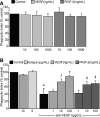
VEGF enhances uptake of apoptotic cells by murine alveolar macrophages. A: AMs were cultured in complete media and pretreated with rmVEGF120 or rmPlGF-2 for 2 h before addition of apoptotic thymocytes. Phagocytic uptake was assessed after 2 h of coculture. Control phagocytic index was 3.7 ± 0.8. N = 2. B: AMs were cultured for 24 h in DMEM with 10% FCS and a VEGF neutralizing antibody. Media was removed, and apoptotic thymocytes were added in serum-free media supplemented with rmVEGF164 or PlGF-2. Phagocytosis was assessed 3 h later. Control phagocytic index was 3.5 ± 0.5. N = 3.*P < 0.05 versus IgG isotype control. †P < 0.05 versus anti-VEGF ab-treated AMs. ‡P < 0.07 versus anti-VEGF ab treated AMs.
Fig. 4.
Fig. 4.
Inhibition of VEGF signaling decreases apoptotic cell uptake by human monocytes-derived macrophages (HMDMs). A: HMDMs were treated with VEGF-depleting antibody or IgG isotype, and cocultured with apoptotic Jurkat cells. Phagocytosis was assessed 1 h later. Control phagocytic index was 20.1 ± 1.1. N = 5. B: HMDMs were treated with SU5416 or DMSO control and cocultured with apoptotic Jurkat cells. Control phagocytic index was 20.9 ± 1.2. N = 5–6. C: HMDMs were treated with VEGF R1 blocking antibody or IgG isotype control and exposed to phosphatidylserine-expressing red blood cells (RBCs). Phagocytosis was assessed 1 h later. Control phagocytic index was 109.7 ± 24.2. N = 4. *P < 0.05 versus isotype control.
Fig. 5.
Fig. 5.
VEGF supplementation enhances Rac1 activation in RAW 264.7 cells. After serum starvation in X-vivo, RAW 264.7 cells were treated with rmVEGF164 (100 ng/ml), LPS-positive control (200 ng/ml), or PBS control. Cells were lysed, harvested, and processed using the Rac1 G-Lisa activation assay. N = 4. *P < 0.05 versus control. ‡P < 0.07 versus control.
Fig. 6.
Fig. 6.
VEGF augmentation enhances apoptotic cell uptake but VEGF receptor inhibition does not decrease efferocytic activity in the murine lung. A: C57BL/6 mice were treated with a single subcutaneous dose of SU5416 or vehicle control. Apoptotic thymocytes were intratracheally instilled 7 days after SU5416 treatment. AM phagocytosis was assessed 1 h later. Control phagocytic index was 5.0 ± 0.5. N ≥ 19 per group. P = 0.12 for SU5416 20 mg/kg dose. B: C57BL/6 mice were treated with intraperitoneal anti-VEGF R1 (800 μg), anti-VEGF R2 (800 μg), both anti-VEGF R1 and anti-VEGF R2, or rat IgG on days 0, 3, and 5. Apoptotic thymocytes were administered intratracheally on day 6. Phagocytosis was assessed in AMs from bronchoalveolar lavage (BAL) 1 h later. Control phagocytic index was 7.1 ± 1.9. N = 6 per group. C and D: transgenic mice with doxycycline (doxy)-inducible expression of human VEGF165 and C57BL/6 mice were treated with doxy-containing or regular chow. At 3 (C) or 7 (D) days after transgene activation, apoptotic thymocytes were instilled intratracheally. AMs were then harvested by BAL 1 h after instillation and phagocytic uptake was assessed. WT, wild-type mice; OE, overexpressor mice. N = 11–12 per group and 5 per group, respectively. *P < 0.05 versus WT/doxy, VEGF OE/Reg, and WT/Reg. ‡P < 0.05 versus WT/Reg.

Similar articles

Cited by

References

    1. Adini A, Kornaga T, Firoozbakht F, Benjamin LE. Placental growth factor is a survival factor for tumor endothelial cells and macrophages. Cancer Res 62: 2749– 2752, 2002 - PubMed
    1. Berse B, Brown LF, Van De Water L, Dvorak HF, Senger DR. Vascular permeability factor (vascular endothelial growth factor) gene is expressed differenetially in normal tissues, macrophages, and tumors. Mol Biol Cell 3: 211– 220, 1992 - PMC - PubMed
    1. Bhandari V. Essential role of nitric oxide in VEGF-induced, asthma-like angiogenic, inflammatory, mucus, and physiologic responses in the lung. Proc Natl Acad Sci USA 103: 11021– 11026, 2006 - PMC - PubMed
    1. Bhandari V, Choo-Wing R, Chapoval SP, Lee CG, Tang C, Kim YK, Ma B, Baluk P, Lin MI, McDonald DM, Homer RJ, Sessa WC, Elias JA. Essential role of nitric oxide in VEGF-induced, asthma-like angiogenic, inflammatory, mucus, and physiologic responses in the lung. Proc Natl Acad Sci USA 103: 11021– 11026, 2006 - PMC - PubMed
    1. Brown KRS, England KM, Goss KL, Snyder JM, Acarregui MJ. VEGF induces airway epithelial cell proliferation in human fetal lung in vitro. Am J Physiol Lung Cell Mol Physiol 281: L1001– L1010, 2001 - PubMed

Publication types

MeSH terms

LinkOut - more resources