. 2012 May;27(5):1004-17.
doi: 10.1002/jbmr.1562.

Multipotent progenitors resident in the skeletal muscle interstitium exhibit robust BMP-dependent osteogenic activity and mediate heterotopic ossification

Affiliations

Multipotent progenitors resident in the skeletal muscle interstitium exhibit robust BMP-dependent osteogenic activity and mediate heterotopic ossification

Michael N Wosczyna et al. J Bone Miner Res. 2012 May.

Abstract

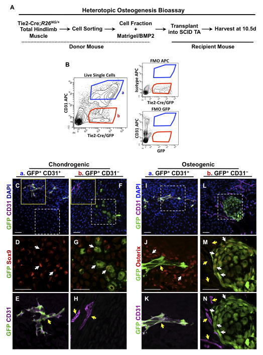
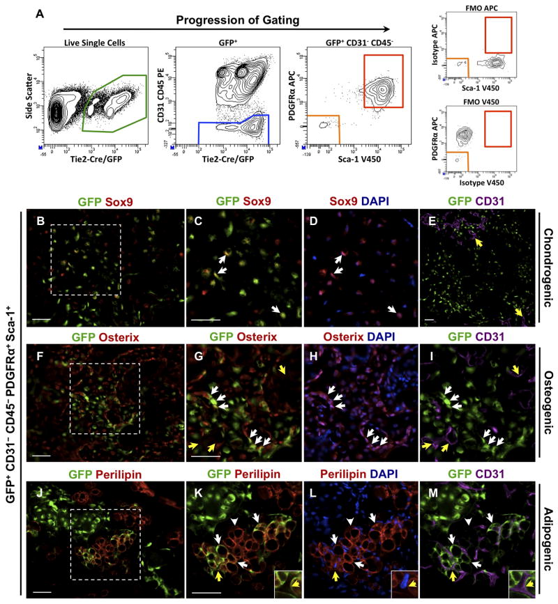
Heterotopic ossification is a debilitating condition that can result from traumatic injury, surgery, or genetic disease. We investigated the cellular origins of heterotopic skeletogenesis in the mouse using lineage tracing and bioassays of heterotopic ossification based on intramuscular transplantation. We identified, characterized, and purified a tissue-resident stem/progenitor cell population that exhibits robust osteogenic potential and represents a major cell-of-origin for heterotopic ossification. These progenitors reside in the interstitium of skeletal muscle and other tissues, and are distinct from the endothelium, which does not exhibit osteogenic activity in response to bone morphogenetic protein 2 (BMP2) stimulation. Intramuscular transplantation, together with clonal analysis in culture, revealed that these progenitors are multipotent, exhibiting the capacity for both BMP-dependent skeletogenic differentiation and spontaneous adipogenic differentiation. Identifying the cells-of-origin responsible for heterotopic ossification provides a potential therapeutic target to treat, mitigate, or prevent this disabling condition.


Figures

Figure 1
Figure 1
Tie2+ cells from a non-endothelial origin contribute to BMP2-induced heterotopic ossification. (A) Lateral view of a mouse hindlimb showing a heterotopic lesion (dashed oval) in the TA muscle 15d after BMP2 administration. (B, C) H&E stained cryostat sections of typical lesions 8d and 15d after BMP2 injection. (B) 8d lesions are primarily comprised of cartilage (C) and surrounding fibroproliferative cells (FP). M, muscle fibers. (C) By 15d, cartilage largely has been replaced by bone (B). Marrow cavities are evident (□). (D–G, L–O) Confocal immunofluorescence images of cartilage and bone stage lesions from Tie2-Cre;R26NG/+ TA muscle. Boxed areas in (D) and (L) are shown at higher magnification in (E, F) and (M–O), respectively. Many GFP+ chondrocytes (Sox9+) and osteogenic cells (Osterix+) are evident (examples shown at white arrows). Examples of GFP+ endothelium are shown at yellow arrows. (H–K, P–S) Sections of representative cartilage and bone stage lesions from VE-Cadherin-Cre;R26NG/+ TA muscle. Boxed areas in (H) and (P) are shown at higher magnification in (I, J) and (Q–S), respectively. CD31+ lesional vasculature is GFP+ (yellow arrows) whereas heterotopic cartilage and bone are GFP-. Images are representative of a minimum of three independent lesions from three mice at each stage. Scale bars are 40μm.
Figure 2
Figure 2
Non-endothelial progenitors participate in heterotopic ossification following intramuscular transplantation. (A) Schematic of the experimental design. (B) After sorting for live, mononuclear cells (Supplemental Fig. S2), GFP+ cells from Tie2-Cre;R26NG/+ total hindlimb muscles were fractionated by flow cytometry into CD31+ (endothelial cells) and CD31- populations and tested for osteogenic activity. Strict sorting gates were applied that minimized cross population contamination and fluorescence-minus-one controls (FMOs) were conducted to assess labeling specificity. ~2 X 10⁴ cells in 50μl were injected in a typical experiment. (C–E, I–K) Confocal images of cryostat sections of heterotopic lesions 10.5d after transplantation of GFP+/CD31+ cells. Solid and dashed boxed areas are shown at higher magnification in (E) and (D, J, K), respectively. GFP+/CD31+ cells contribute to CD31+ lesional vasculature (yellow arrows), but not to Sox9+ cartilage or Osterix+ bone of heterotopic lesions (white arrows). (F–H, L–N) GFP+/CD31- cells contribute to chondrogenic and osteogenic cells of the skeletal anlagen (white arrows) but do not contribute to endothelium (yellow arrows). Solid and dashed boxed areas are shown at higher magnification in (H) and (G, M, N), respectively. Images are representative of a minimum of two independent lesions at each stage. Scale bars are 40μm.
Figure 3
Figure 3
Identification of GFP+CD31-CD45-PDGFRα+Sca-1+ skeletogenic progenitors from uninjured skeletal muscle. (A) After sorting for live, mononuclear cells (Supplemental Fig. S2), GFP+ cells from Tie2-Cre;R26NG/+ total hindlimb muscles were fractionated by flow cytometry. Strict gates were set to minimize cross contamination between populations, and FMO controls were conducted to assess labeling specificity. (B–I) GFP+CD31-CD45-PDGFRα+Sca-1+ cells were tested for osteogenic activity as in Fig. 2. Boxed areas in (B) and (F) are shown at higher magnification in (C, D) and (G–I), respectively. The contribution of GFP+ cells to chondrocytes and osteoblasts/osteocytes of induced lesions was readily apparent (white arrows), based on Sox9 and Osterix staining, respectively. Endothelium was GFP- (yellow arrows in E, G, I). (J–M) Donor-derived Perilipin+ adipocytes were consistently observed (white arrows). Adipocytes typically were unilocular and did not stain with the brown fat marker UCP1, indicating a white fat phenotype. GFP- host-derived adipocytes were also apparent (arrowheads in K–M). Insets in (K–M) are high magnification views of the area noted by the yellow arrows, showing the distinction between GFP+ adipocytes and GFP-CD31+ endothelium. Images are representative of a minimum of four independent lesions for each stage. Scale bars are 50μm.
Figure 4
Figure 4
Cell surface antigen expression of GFP+CD31-CD45-PDGFRα+Sca-1+ progenitors derived from Tie2-Cre;R26NG/+ skeletal muscle. (A–C) Among GFP+CD31-CD45- cells, ~96% of Sca-1+ cells also express PDGFRα+, whereas ~99% of PDGFRα+ cells express Sca-1. Therefore, expression of other surface markers was assessed by replacing the anti-PDGFRα+ APC antibody (A, B) or the anti-Sca-1 V450 antibody (C) with antibodies to other markers, as shown. Quadrants were established based on FMO controls. Percentages in each quadrant represent the proportion of total GFP+CD31-CD45- cells.
Figure 5
Figure 5
Clonal analysis of GFP+CD31-CD45-PDGFRα+Sca-1+ progenitors in culture. (A) Single cells were sorted directly into individual wells of 96 well plates and evaluated for adipogenic and osteogenic differentiation, with or without the addition of BMP2 on day 14 of culture. (B, C) Typical example of a live colony maintained in growth medium for 12d. Insets are magnified views of the boxed areas. Adipocytes with multilocular lipid deposits are apparent (insets). Panel B, Hoffman modulation contrast microscopy. (D) 14d culture stained with Oil Red O to definitively identify lipid droplets. (E–H) Typical examples of 23d cultures maintained in growth medium and stained for the osteogenic markers, ALP (E) or Osterix (F, G), or for the adipogenic marker, Perilipin (H). 89% of colonies were negative for ALP activity, and no cells with nuclear localized Osterix immunoreactivity were observed. Perilipin+ adipocytes were abundant in these cultures. Fibroblastic-like cells that stain for SMA are also present in these cultures (data not shown). (I–L) Examples of 23d cultures that were switched from growth medium to BMP2-containing osteogenic medium on day 14. Most colonies were characterized by intense ALP staining (I) or nuclear localized Osterix expression (J, K; arrows). Perilipin+ adipogenic cells persisted under osteogenesis-inducing conditions (L). Scale bars represent 100μm (B, D, E, I), or 50μm (F, J, H, L). (M) Quantification of adipogenic and osteogenic differentiation in clonal cultures. Adipogenic differentiation was assessed between 10 and 14 days of culture, and colonies were scored as positive if they contained cells with large lipid droplets, stained with Oil Red O, or expressed Perilipin (representative examples of positive criteria are shown in panels B, D, H, and I. Colonies were scored as osteogenic if they contained cells that stained positively for either ALP activity or nuclear-localized Osterix expression. Osterix was not detected in growth media cultures. Error bars represent ± SEM. Data is from three independent experiments. *P< 0.001 (Chi-Square Test).
Figure 6
Figure 6
GFP+CD31-CD45-PDGFRα+Sca-1+ progenitors reside in the skeletal muscle interstitium of Tie2-Cre;R26NG/+ mice. The Soleus muscle is shown. Results with the EDL and TA muscles were comparable. (A) Confocal image of a muscle cross section that was immunostained for Sca-1 and PDGFRα. Dotted lines represent muscle fiber borders. A GFP+ cell that expresses both PDGFRα and Sca-1 is visible in this view (white arrow and lower right insets). Note its close association with the adjacent GFP+Sca-1+ endothelial cell (yellow arrow; also see panel B). Two GFP- cells that express PDGFRα+ and are weakly positive for Sca-1 are also present (purple arrows). Lower left insets: cell at smaller purple arrows. Levels for the red channel were increased for the left inset to visualize the faint Sca-1 staining. (B) GFP+PDGFRα+ cells (white arrows and insets) were often observed in close proximity to CD31+ endothelium (yellow arrow and insets). Note that ~99% of GFP+PDGFRα+ cells were also positive for Sca-1 (Fig. 4A). (C) GFP+PDGFRα+ cells (white arrows and insets) do not share a basal lamina (Laminin+) with endothelial cells (yellow arrows and insets), indicating that these progenitor cells are distinct from pericytes. GFP+PDGFRα+ cells reside in interstitial spaces between muscle fibers, each of which is circumscribed by a Laminin+ basal lamina. (D) GFP+PDGFRα+ cells (white arrows and inset) do not express the pericyte marker, NG2 (blue arrows). Images are representative of sections taken from a minimum of two Soleus muscles isolated from two independent mice. Scale bars are 20μm.

Similar articles

Cited by

References

    1. Cushner FD, Morwessel RM. Myositis ossificans traumatica. Orthop Rev. 1992;21 (11):1319–26. - PubMed
    1. Potter BK, Burns TC, Lacap AP, Granville RR, Gajewski DA. Heterotopic ossification following traumatic and combat-related amputations. Prevalence, risk factors, and preliminary results of excision. J Bone Joint Surg Am. 2007;89(3):476–86. - PubMed
    1. Shore EM, Kaplan FS. Insights from a rare genetic disorder of extra-skeletal bone formation, fibrodysplasia ossificans progressiva (FOP) Bone. 2008;43(3):427–33. - PMC - PubMed
    1. Potter BK, Burns TC, Lacap AP, Granville RR, Gajewski D. Heterotopic ossification in the residual limbs of traumatic and combat-related amputees. J Am Acad Orthop Surg. 2006;14(10 Suppl):S191–7. - PubMed
    1. Vanden Bossche L, Vanderstraeten G. Heterotopic ossification: a review. J Rehabil Med. 2005;37(3):129–36. - PubMed

Publication types