. 2012 Mar 23;287(13):10631-10638.
doi: 10.1074/jbc.M111.299917. Epub 2012 Feb 3.

Autophagy-related protein 32 acts as autophagic degron and directly initiates mitophagy

Affiliations

Autophagy-related protein 32 acts as autophagic degron and directly initiates mitophagy

Noriko Kondo-Okamoto et al. J Biol Chem. .

Abstract

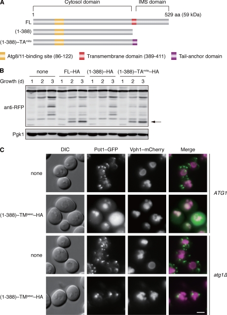
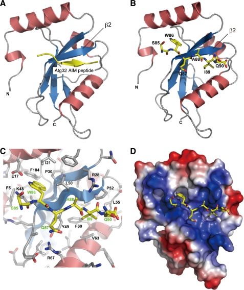
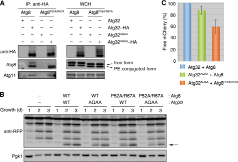
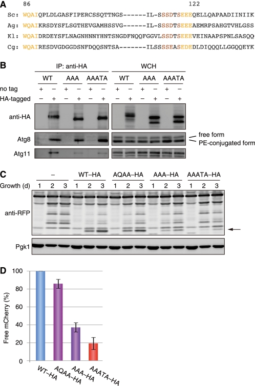
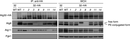
Autophagy-related degradation selective for mitochondria (mitophagy) is an evolutionarily conserved process that is thought to be critical for mitochondrial quality and quantity control. In budding yeast, autophagy-related protein 32 (Atg32) is inserted into the outer membrane of mitochondria with its N- and C-terminal domains exposed to the cytosol and mitochondrial intermembrane space, respectively, and plays an essential role in mitophagy. Atg32 interacts with Atg8, a ubiquitin-like protein localized to the autophagosome, and Atg11, a scaffold protein required for selective autophagy-related pathways, although the significance of these interactions remains elusive. In addition, whether Atg32 is the sole protein necessary and sufficient for initiation of autophagosome formation has not been addressed. Here we show that the Atg32 IMS domain is dispensable for mitophagy. Notably, when anchored to peroxisomes, the Atg32 cytosol domain promoted autophagy-dependent peroxisome degradation, suggesting that Atg32 contains a module compatible for other organelle autophagy. X-ray crystallography reveals that the Atg32 Atg8 family-interacting motif peptide binds Atg8 in a conserved manner. Mutations in this binding interface impair association of Atg32 with the free form of Atg8 and mitophagy. Moreover, Atg32 variants, which do not stably interact with Atg11, are strongly defective in mitochondrial degradation. Finally, we demonstrate that Atg32 forms a complex with Atg8 and Atg11 prior to and independent of isolation membrane generation and subsequent autophagosome formation. Taken together, our data implicate Atg32 as a bipartite platform recruiting Atg8 and Atg11 to the mitochondrial surface and forming an initiator complex crucial for mitophagy.


Figures

FIGURE 1.
FIGURE 1.
Atg32 contains a domain compatible for degradation of peroxisomes. A, schematic representation of the domain structures of Atg32 (FL) and its truncated variants. (1–388) lacks the transmembrane and IMS domains, and (1–388) -TAmito consists of the cytosolic domain and a tail-anchor derived from Gem1, a mitochondrial outer membrane protein (39). B, cells expressing the wild-type or truncated variants of HA-tagged Atg32 (FL-HA, (1–388)–HA, or (1–388)-TAmito-HA) were grown in glycerol medium, collected at the indicated time points, and subjected to Western blotting. All strains are atg32-null derivatives expressing a mitochondrial matrix-localized DHFR-mCherry. The arrow depicts free mCherry generated by mitophagy. Pgk1 was monitored as a loading control. C, cells containing or lacking Atg1 (ATG1 or atg1Δ) were transformed with a low-copy, empty plasmid (none) or the one that encodes (1–388)-TMpexo-HA, an Atg32 cytosol domain anchored to the peroxisome via a TM domain derived from Pex15, a peroxisomal membrane protein (40). (1–388)-TMpexo-HA was expressed under the ATG32 promoter. All strains are atg32-null derivatives expressing both Pot1-GFP (peroxisomal marker) and Vph1-mCherry (vacuolar marker). Cells were grown in glycerol medium for 48 h and analyzed by fluorescence microscopy. Scale bar = 2 μm.
FIGURE 2.
FIGURE 2.
Crystal structure of the Atg8-Atg32(SWQAIQ)85–90 peptide complex. A, Atg8 (α helices in red, β sheets in blue) and the Atg32 peptide (yellow) are shown in ribbon models. B, Atg8 and the Atg32(SWQAIQ)85–90 peptide are shown in ribbon and stick models, respectively. C, close-up view of the Atg8-Atg32(SWQAIQ)85–90 interface indicating amino acids of Atg8 (black) and Atg32 (green). D, Atg8 and the Atg32(SWQAIQ)85–90 peptide are shown in surface and stick models, respectively. The surface is colored according to the electrostatic potential (blue, positive; red, negative).
FIGURE 3.
FIGURE 3.
Atg32-Atg8 interaction contributes to efficient mitophagy. A, coimmunoprecipitation assays for cells expressing the indicated variants of Atg8 and Atg32 grown in glycerol medium for 30 h. All strains are vacuolar protease-deficient, atg8- and atg32-double null derivatives. Mitochondria-enriched fractions were obtained from whole cell homogenates (WCH), solubilized, and subjected to immunoprecipitation using anti-HA antibody-conjugated agarose. The WCH fractions and eluted immunoprecipitates (IP) were analyzed by Western blotting. B, cells expressing the wild-type or mutants of Atg8 (WT or P52A/R67A) and Atg32 (WT or AQAA) were grown in glycerol medium, collected at the indicated time points, and subjected to Western blotting. All strains are atg8- and atg32-double-null derivatives expressing a mitochondrial matrix-localized DHFR-mCherry. The arrow depicts free mCherry generated by mitophagy. Pgk1 was monitored as a loading control. C, the amounts of free mCherry generated in cells expressing the indicated variants of Atg32 and Atg8 at the 3-day time point were analyzed as in B and quantified in three experiments. The signal intensity value of free mCherry in cells expressing wild-type Atg32-HA and Atg8 was set to 100%. Data represent the averages of the all experiments, with bars indicating mean ± S.D.
FIGURE 4.
FIGURE 4.
Atg32-Atg11 interaction is crucial for mitochondria autophagy. A, an amino acid sequence alignment of the regions containing putative Atg8- and Atg11-binding domains from yeast Atg32 homologs. Sc, S. cerevisiae; Ag, Ashbya gossypii; Kl, Kluyveromyces lactis; Cg, Candida glabrata. B, coimmunoprecipitation assays for cells expressing untagged and HA-tagged variants of Atg32 (WT), Atg32AAA (AAA) for EEE120–122, or Atg32AAATA (AAATA) for SSDTS115–119 grown in glycerol medium for 30 h. All strains are vacuolar protease-deficient, atg32-null derivatives. Mitochondria-enriched fractions were obtained from whole cell homogenates (WCH), solubilized, and subjected to immunoprecipitation using anti-HA antibody-conjugated agarose. The WCH fractions and eluted immunoprecipitates (IP) were analyzed by Western blotting. C, cells expressing the wild-type or mutants of HA-tagged Atg32 (WT-HA, AQAA-HA, AAA-HA, or AAATA-HA) were grown in glycerol medium, collected at the indicated time points, and subjected to Western blotting. All strains are atg32-null derivatives expressing a mitochondrial matrix-localized DHFR-mCherry. The arrow depicts free mCherry generated by mitophagy. Pgk1 was monitored as a loading control. D, the amounts of free mCherry generated in cells expressing the indicated variants of Atg32 at the 3-day time point were analyzed as in C and quantified in three experiments. The signal intensity value of free mCherry in cells expressing wild-type Atg32-HA was set to 100%. Data represent the averages of the all experiments, with bars indicating mean ± S.D.
FIGURE 5.
FIGURE 5.
Atg32-Atg8 and -Atg11 interactions occur before isolation membrane generation. Coimmunoprecipitation assays for autophagy-competent (WT) and atg114 null mutant (1Δ∼14Δ) cells expressing Atg32 (32) or Atg32-HA (32-HA) grown in glycerol medium for 30 h. All strains are vacuolar protease-deficient, atg32-null derivatives. Mitochondria-enriched fractions were obtained from whole cell homogenates (WCH), solubilized, and subjected to immunoprecipitation using anti-HA antibody-conjugated agarose. The WCH fractions and eluted immunoprecipitates (IP) were analyzed by Western blotting.

Similar articles

Cited by

References

    1. Weidberg H., Shvets E., Elazar Z. (2011) Biogenesis and cargo selectivity of autophagosomes. Annu. Rev. Biochem. 80, 125–156 - PubMed
    1. Mizushima N., Yoshimori T., Ohsumi Y. (2011) Annu. Rev. Cell Dev. Biol. 27, 107–132 - PubMed
    1. Youle R. J., Narendra D. P. (2011) Mechanisms of mitophagy. Nat. Rev. Mol. Cell Biol. 12, 9–14 - PMC - PubMed
    1. Okamoto K., Kondo-Okamoto N. (2012) Biochim. Biophys. Acta in press - PubMed
    1. Zhang J., Ney P. A. (2011) Mechanisms and biology of B-cell leukemia/lymphoma 2/adenovirus E1B-interacting protein 3 and Nip-like protein X. Antioxid. Redox Signal. 14, 1959–1969 - PMC - PubMed

Publication types

MeSH terms

Substances