. 2012 Mar 23;287(13):10664-10673.
doi: 10.1074/jbc.M111.320929. Epub 2012 Feb 3.

Selenocysteine insertion sequence (SECIS)-binding protein 2 alters conformational dynamics of residues involved in tRNA accommodation in 80 S ribosomes

Affiliations

Selenocysteine insertion sequence (SECIS)-binding protein 2 alters conformational dynamics of residues involved in tRNA accommodation in 80 S ribosomes

Kelvin Caban et al. J Biol Chem. .

Abstract

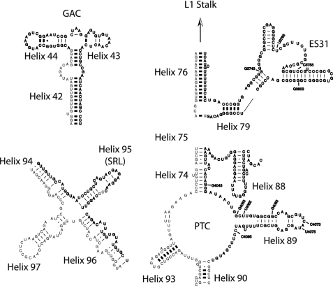
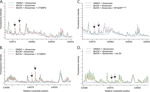
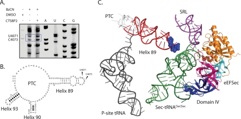
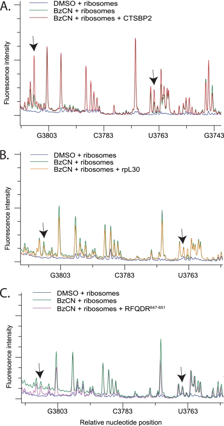
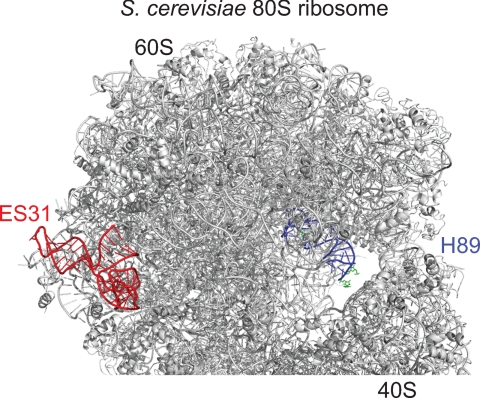
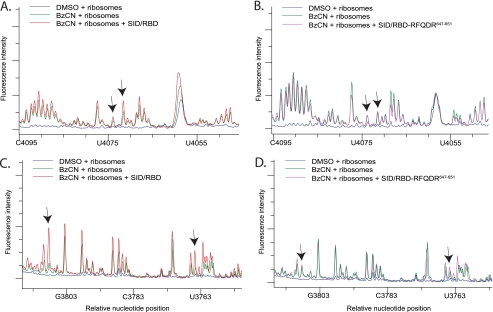
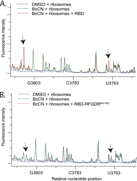
Sec-tRNA(Sec) is site-specifically delivered at defined UGA codons in selenoprotein mRNAs. This recoding event is specified by the selenocysteine insertion sequence (SECIS) element and requires the selenocysteine (Sec)-specific elongation factor, eEFSec, and the SECIS binding protein, SBP2. Sec-tRNA(Sec) is delivered to the ribosome by eEFSec-GTP, but this ternary complex is not sufficient for Sec incorporation, indicating that its access to the ribosomal A-site is regulated. SBP2 stably associates with ribosomes, and mutagenic analysis indicates that this interaction is essential for Sec incorporation. However, the ribosomal function of SBP2 has not been elucidated. To shed light on the functional relevance of the SBP2-ribosome interaction, we screened the functional centers of the 28 S rRNA in translationally competent 80 S ribosomes using selective 2'-hydroxyl acylation analyzed by primer extension (SHAPE). We demonstrate that SBP2 specifically alters the reactivity of specific residues in Helix 89 (H89) and expansion segment 31 (ES31). These results are indicative of a conformational change in response to SBP2 binding. Based on the known functions of H89 during translation, we propose that SBP2 allows Sec incorporation by either promoting Sec-tRNA(Sec) accommodation into the peptidyltransferase center and/or by stimulating the ribosome-dependent GTPase activity of eEFSec.


Figures

FIGURE 1.
FIGURE 1.
Mouse 28 S rRNA. The reactivity of bases found at or near the GAC, the SRL, the PTC, and ES31 indicated in bold face type were interrogated with BzCN. The approximate base positions shown in the electrophoretic traces in Figs. 2, 4, 6, and 7 are also indicated. The secondary structures were adapted from the mouse 28 S rRNA structure obtained in the Comparative RNA web site and project.
FIGURE 2.
FIGURE 2.
CTSBP2 specifically enhances the reactivity of nucleotides in H89. A, electrophoretic traces obtained from reactions containing 80S ribosomes treated with 80 mm BzCN in the presence (red) or absence (green) of recombinant CT-SBP2, or DMSO (blue). cDNA fragments were generated from primer extension reactions using primer 4168. B, same as in A except primer extension was performed with a second primer (primer 4229) located further downstream (see “Experimental Procedures”). C, Same as A except reactions were performed with the CTSBP2 647RFQDR651 penta-alanine mutant (purple). D, same as B except reactions were performed with ribosomal protein L30 (rpL30, orange). The y axis shows the relative fluorescence intensity, and the x axis shows the approximate nucleotide position on the mouse 28 S rRNA. Arrows indicate the nucleotides displaying enhanced reactivity in the presence of CTSBP2 (A and B) or the corresponding positions in reactions performed with the CTSBP2 647RFQDR651 penta-alanine mutant (C) or rpL30 (D). Each trace is representative of at least three independent SHAPE experiments.
FIGURE 3.
FIGURE 3.
Localization of SBP2-induced modifications in H89. A, sequencing lanes are indicated by A, U, C, or G and were generated from rRNA extracted from ribosomes. Primer extension reactions were generated from rRNA extracted from ribosomes incubated with DMSO, or BzCN in the presence or absence of CTSBP2. Residues U4071 and C4073 (E. coli U2473 and C2475, respectively), which display enhanced reactivity in the presence of CTSBP2, are indicated by the blue box. B, mouse 28 S secondary structure map of H89 with bases U4071 and C4073 indicated. C, PyMOL-generated image of the eEFSec homolog, archaeal SelB (Protein Data Bank code 1WB3), and the human tRNASec (Protein Data Bank code 3A3A), aligned over the crystal structure of the EF-Tu TC bound to the 70 S ribosome (2WRQ and 2WRR) using the align command. Domain I (orange), II (teal), III (magenta), and IV (blue) of eEFSec are also highlighted. H89, which is projected from the PTC (white), is colored in red, and residues U2473 and C2475 are shown as blue spheres. The P-site tRNA (dark gray) and the SRL (purple) are also indicated for reference.
FIGURE 4.
FIGURE 4.
CTSBP2 specifically enhances the reactivity of bases in ES31. A, electrophoretic traces obtained from reactions containing 80 S ribosomes treated with DMSO (blue), or BzCN in the presence (red) or absence (green) of CTSBP2. cDNA fragments were generated from primer extension reactions using primer 3928. B and C, same as A except reactions were performed with the CTSBP2 647RFQDR651 penta-alanine mutant (purple) or rpL30 (orange). Arrows indicate regions with enhanced reactivity in the presence of CTSBP2 (A), or the corresponding positions in reactions performed with the CTSBP2 647RFQDR651 penta-alanine mutant (B) or rpL30 (C). Each trace is representative of three independent SHAPE experiments.
FIGURE 5.
FIGURE 5.
Relative positions of ES31(red) and H89 (blue) on the crystal structure of the S. cerevisiae 80 S ribosome. The residues shown in green on H89 displayed enhanced reactivity to BzCN in the presence of SBP2.
FIGURE 6.
FIGURE 6.
SHAPE analysis of H89 and ES31 with the SBP2 SID/RBD domains. A and C, electrophoretic traces obtained from reactions containing ribosomes treated with DMSO (blue) or BzCN in the presence (red) or absence (green) of the SID and the RBD. cDNA fragments were generated from reactions using primer 4229 to cover H89 (A) and primer 3928 to cover ES31 (C). B and D, same as A and C except reactions were performed with the SID and the RBD 647RFQDR651 penta-alanine mutant (purple). Arrows in A, B and C, D indicate the same nucleotide positions displaying enhanced reactivity in the presence of CTSBP2 seen in Figs. 2B and 3A, respectively. Each trace is representative of three independent SHAPE experiments.
FIGURE 7.
FIGURE 7.
SHAPE analysis of ES31 with the SBP2 RBD domain alone. A, electrophoretic traces obtained from reactions containing ribosomes treated with DMSO (blue), or BzCN in the presence (red) or absence (green) of the RBD. cDNA fragments were generated from reactions using primer 3928 to cover ES31. B, same as A except reactions were performed with the RBD 647RFQDR651 penta-alanine mutant (purple). Arrows indicate the same nucleotide positions displaying enhanced reactivity in the presence of CTSBP2 seen in Fig. 3A. Each trace is representative of three independent SHAPE experiments.

Similar articles

Cited by

References

    1. Fagegaltier D., Hubert N., Yamada K., Mizutani T., Carbon P., Krol A. (2000) Characterization of mSelB, a novel mammalian elongation factor for selenoprotein translation. EMBO J. 19, 4796–4805 - PMC - PubMed
    1. Tujebajeva R. M., Copeland P. R., Xu X. M., Carlson B. A., Harney J. W., Driscoll D. M., Hatfield D. L., Berry M. J. (2000) Decoding apparatus for eukaryotic selenocysteine insertion. EMBO Rep. 1, 158–163 - PMC - PubMed
    1. Diamond A., Dudock B., Hatfield D. (1981) Structure and properties of a bovine liver UGA suppressor serine tRNA with a tryptophan anticodon. Cell 25, 497–506 - PubMed
    1. Jung J. E., Karoor V., Sandbaken M. G., Lee B. J., Ohama T., Gesteland R. F., Atkins J. F., Mullenbach G. T., Hill K. E., Wahba A. J. (1994) Utilization of selenocysteyl-tRNA[Ser]Sec and seryl-tRNA[Ser]Sec in protein synthesis. J. Biol. Chem. 269, 29739–29745 - PubMed
    1. Fahlman R. P., Dale T., Uhlenbeck O. C. (2004) Uniform binding of aminoacylated transfer RNAs to the ribosomal A and P sites. Mol. Cell 16, 799–805 - PubMed

Publication types

MeSH terms

LinkOut - more resources