. 2012 Mar 30;287(14):10922-32.
doi: 10.1074/jbc.M111.333393. Epub 2012 Feb 3.

Structural dynamics of bacterial translation initiation factor IF2

Affiliations

Structural dynamics of bacterial translation initiation factor IF2

Hans Wienk et al. J Biol Chem. .

Abstract

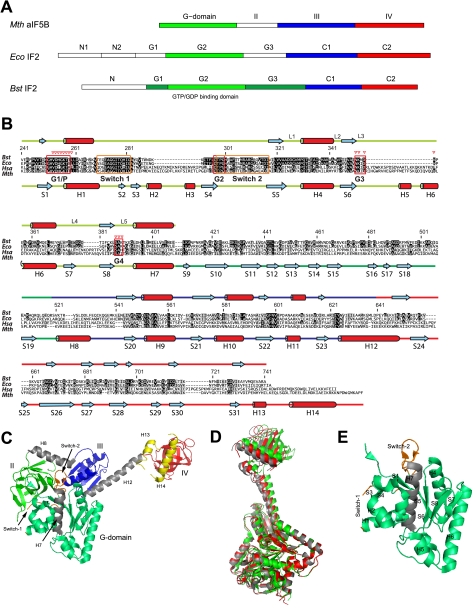
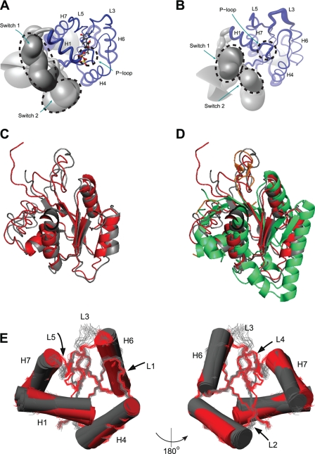
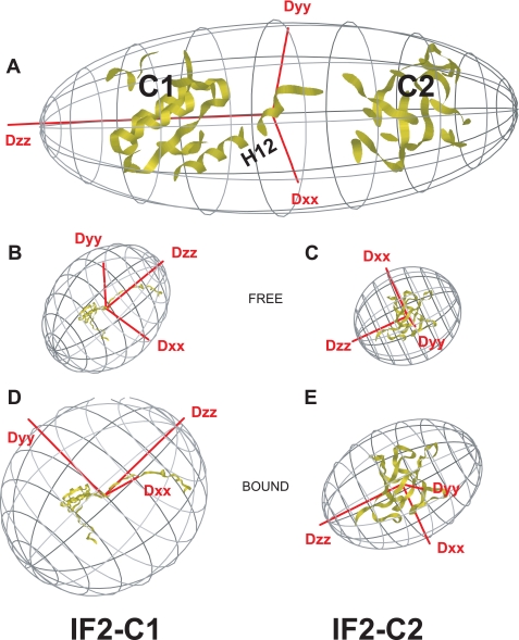
Bacterial translation initiation factor IF2 promotes ribosomal subunit association, recruitment, and binding of fMet-tRNA to the ribosomal P-site and initiation dipeptide formation. Here, we present the solution structures of GDP-bound and apo-IF2-G2 of Bacillus stearothermophilus and provide evidence that this isolated domain binds the 50 S ribosomal subunit and hydrolyzes GTP. Differences between the free and GDP-bound structures of IF2-G2 suggest that domain reorganization within the G2-G3-C1 regions underlies the different structural requirements of IF2 during the initiation process. However, these structural signals are unlikely forwarded from IF2-G2 to the C-terminal fMet-tRNA binding domain (IF2-C2) because the connected IF2-C1 and IF2-C2 modules show completely independent mobility, indicating that the bacterial interdomain connector lacks the rigidity that was found in the archaeal IF2 homolog aIF5B.


Figures

FIGURE 1.
FIGURE 1.
Domain organization of IF2 and aIF5B. A, domain nomenclature for IF2 homologs from M. thermoautotrophicum (Mth), E. coli (Eco), and B. stearothermophilus (Bst). B, sequence alignment for IF2 homologs from B. stearothermophilus (Bst), E. coli (Eco), Homo sapiens (Hsa), and M. thermoautotrophicum (Mth). Amino acid numbering is for B. stearothermophilus IF2. More and less conserved residues are depicted in black and gray, respectively. Below the sequence alignment the helices (H) and β-strands (S) found in M. thermoautotrophicum aIF5B crystal structures are indicated, connected by colored lines, representing IF2-G2 (light green), IF2-G3 (dark green), IF2-C1 (blue), and IF2-C2 (red). Above the sequence alignment the secondary structure elements as found by NMR on B. stearothermophilus IF2 are shown. Guanine nucleotide binding boxes G1–G4, loops L1-L5, and Switch-1 and Switch-2 regions are indicated; red triangles indicate residues that contact the ligand. C, M. thermoautotrophicum aIF5B domain organization. Domain-connecting helices H7, H8, and H12 are colored gray; Switch-1 and Switch-2 are colored orange. Two C-terminal a/eIF5B helices H13 and H14, not occurring in B. stearothermophilus IF2, are colored yellow. D, crystal structures for the free (gray), the GDP-bound (red), and the GDPNP-bound (green) states of M. thermoautotrophicum aIF5B fit on the G2 domain backbone (excluding the Switch-1 and Switch-2 regions). E, secondary structure elements of the M. thermoautotrophicum aIF5B-G2 domain.
FIGURE 2.
FIGURE 2.
IF2 binds GDP through a direct hydrogen bond. The figure shows the data of the direct detection of intermolecular JNP scalar couplings. One-dimensional 31P–15N spin-echo difference 15N-HSQC spectrum of IF2-G2·GDP and a schematic view of the phosphate-amide proton hydrogen bonds are detectable with the pulse sequence of supplemental Fig. S2.
FIGURE 3.
FIGURE 3.
Structures of GDP-bound and apo-IF2-G2. A, “sausage” representation of the IF2-G2·GDP NMR ensemble. The thickness of the backbone represents the r.m.s.d. Structural elements are indicated; GDP is shown in “ball-and-stick.” B, sausage representation of apo-IF2-G2. C, backbone overlay of the best structures of the NMR ensembles calculated for apo- (gray) and GDP-bound (red) IF2-G2. D, as C, additionally overlaid with the crystal structure of the G2 domain from M. thermoautotrophicum aIF5B (green). E, different orientations of the reorganization within the B. stearothermophilus IF2-G2 domain between its apo-state (gray) and GDP-bound (red) state.
FIGURE 4.
FIGURE 4.
Hydrodynamic analysis of B. stearothermophilus IF2 C1-C2. The figure shows the orientations of the rotational diffusion tensors for free and connected B. stearothermophilus IF2 C1-C2 domains. A, predicted rotational diffusion tensor for the M. thermoautotrophicum aIF5B-based homology model for B. stearothermophilus IF2 C1-C2 assuming a completely rigid linker. B, rotational diffusion tensor for free IF2-C1 using the B. stearothermophilus IF2-C1 NMR structure. C, rotational diffusion tensor for free IF2-C2 using the B. stearothermophilus IF2-C2 NMR structure. D, rotational diffusion tensor for IF2-C1 when bound to IF2-C2, using the B. stearothermophilus IF2-C1 NMR structure. E, rotational diffusion tensor for IF2-C2 when bound to IF2-C1, using the B. stearothermophilus IF2-C2 NMR structure. Note: for direct comparison, the individual domains were manually rotated to the coordinate frame of IF2 C1-C2 in A. Also, the rotational diffusion tensors are scaled with respect to their rotational correlation times.
FIGURE 5.
FIGURE 5.
Model for the positional mobility of B. stearothermophilus IF2-C2. The figure shows a backbone representation of B. stearothermophilus IF2 C1-C2. Domain connector “H12” (Arg622–Lys646) is shown in green, and residues belonging to IF2-C1 are blue, and residues forming IF2-C2 are in red. The G23 domains are shown (in gray) based on sequence homology and crystal structures of M. thermoautotrophicum aIF5B. Arrows indicate the motional freedom of the C2 domain compared with the rest of the protein. The inset shows the structural changes detected for IF2-G2 upon GDP binding.

References

    1. Boelens R., Gualerzi C. O. (2002) Structure and function of bacterial initiation factors. Curr. Protein Pept. Sci. 3, 107–119 - PubMed
    1. Laursen B. S., Sørensen H. P., Mortensen K. K., Sperling-Petersen H. U. (2005) Initiation of protein synthesis in bacteria. Microbiol. Mol. Biol. Rev. 69, 101–123 - PMC - PubMed
    1. Gualerzi C. O., Fabbretti A., Brandi L., Milon P., Pon C. L. (2010) Role of the initiation factors in mRNA start site selection and fMet-tRNA recruitment by bacterial ribosomes. Isr. J. Chem. 50, 80–94
    1. Milon P., Carotti M., Konevega A. L., Wintermeyer W., Rodnina M. V., Gualerzi C. O. (2010) The ribosome-bound initiation factor 2 recruits initiator tRNA to the 30 S initiation complex. EMBO Rep. 11, 313–316 - PMC - PubMed
    1. Antoun A., Pavlov M. Y., Tenson T., Ehrenberg M. (2004) Ribosome formation from subunits studied by stopped-flow and Rayleigh light scattering. Biol. Proced. Online 6, 35–54 - PMC - PubMed

Publication types

MeSH terms

LinkOut - more resources