. 2012 Mar 16;287(12):9568-78.
doi: 10.1074/jbc.M111.296954. Epub 2012 Feb 3.

Identification of mechanism that couples multisite phosphorylation of Yes-associated protein (YAP) with transcriptional coactivation and regulation of apoptosis

Affiliations

Identification of mechanism that couples multisite phosphorylation of Yes-associated protein (YAP) with transcriptional coactivation and regulation of apoptosis

Kyung-Kwon Lee et al. J Biol Chem. .

Erratum in

Abstract

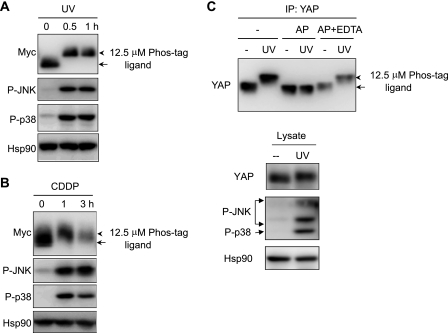
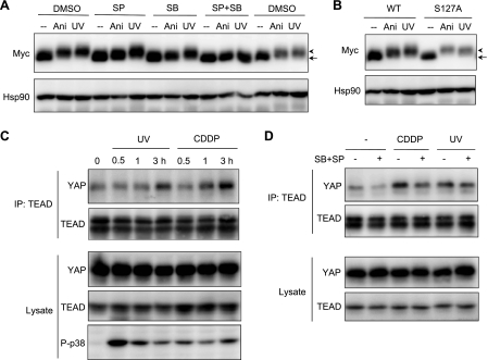
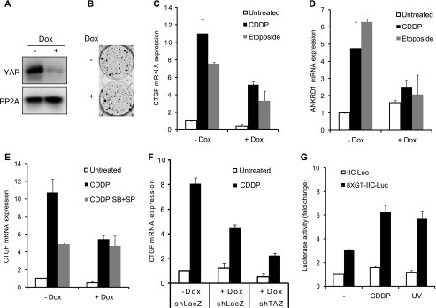
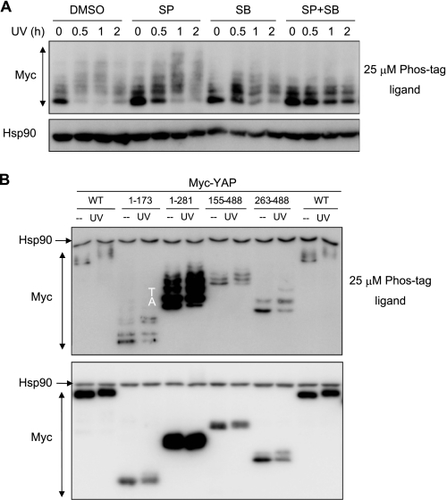
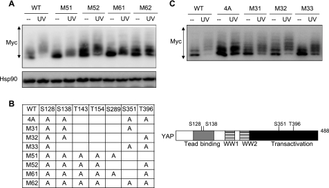
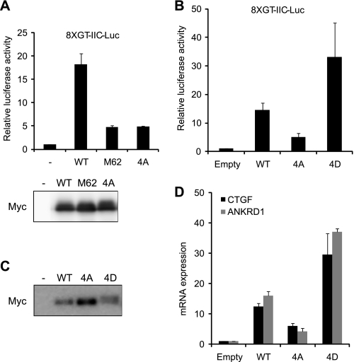
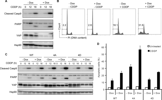
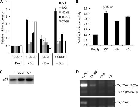
The transcriptional coactivator Yes-associated protein (YAP) has been implicated in tumorigenesis by regulating cell proliferation and apoptosis. YAP interacts with the transcription factor TEAD and is essential in mediating TEAD-dependent gene expression. Here we show that YAP is hyperphosphorylated and activated in response to genotoxic stress such as UV irradiation and cisplatin treatment. Using high resolution mobility shift assay for phosphorylated proteins, we identified multiple sites of phosphorylation induced by UV irradiation. Pretreatment with p38 and JNK inhibitors completely suppressed the mobility retardation of phosphorylated YAP in UV-irradiated cells. Co-immunoprecipitation experiments showed that the physical interaction of YAP with TEAD was markedly enhanced by UV irradiation or CDDP treatment but suppressed by pretreatment with p38 and JNK inhibitors. Similarly, pretreatment with p38 and JNK inhibitors suppressed the expression of YAP/TEAD target genes, which were elevated on exposure to genotoxic stress. Using phosphomimetic and phosphorylation-deficient YAP mutants, we showed that the coactivator activity of YAP correlated with its state of phosphorylation and sensitivity to cisplatin-induced apoptosis. Our results demonstrate that multisite phosphorylation of YAP induces YAP/TEAD-dependent gene expression and provides a mechanism by which YAP regulates apoptosis differently depending on cellular context.


Figures

FIGURE 1.
FIGURE 1.
UV irradiation or CDDP-induced mobility shift of YAP. A and B, 293T cells were transfected with Myc-YAP construct, and cells were UV-irradiated (100 J/m2) (A) or treated with CDDP (100 μm) (B) for the indicated times. Cell lysates were analyzed by immunoblotting with the antibodies indicated. C, YAP was immunoprecipitated with anti-YAP antibody from UV-irradiated KB cell lysates. Immunoprecipitates (IP) were incubated in the absence or presence of calf intestinal alkaline phosphatase (AP) together with or without EDTA (50 mm) and immunoblotted with anti-YAP antibody. The arrows indicate the position of YAP in untreated sample, and arrowheads indicate YAP with slow mobility. Hsp90 was blotted as a loading control. For detection of mobility shift of YAP, cell lysates were resolved in polyacrylamide gels containing 12.5 μm Phos-tag ligand when indicated.
FIGURE 2.
FIGURE 2.
p38 and JNK inhibitors suppress UV irradiation- or CDDP-induced interaction of YAP with TEAD. A, U2OS cells were transfected with Myc-YAP and preincubated for 2 h without (DMSO) or with JNK inhibitor, SP600125 (SP) (10 μm) and/or with p38 inhibitor, SB202190 (SB) (10 μm). Then cells were treated with anisomycin (Ani) (50 μg/ml) or UV-irradiated (100 J/m2). Cell lysates were analyzed by immunoblotting. B, U2OS cells expressing wild-type YAP (WT) or YAP 127A (127A) were treated and analyzed as in A. Immunoblot with anti-Myc antibody was performed on a gel containing 12.5 μm of Phos-tag ligand (A and B). C, KB cells were UV-irradiated or treated with CDDP (100 μm) for 3 h. Cell lysates were immunoprecipitated with anti-TEAD antibody, and coimmunoprecipitates (IP) were analyzed by immunoblotting with indicated antibodies. D, KB cells were preincubated in the absence or presence of p38/JNK inhibitors (SB+SP) for 1 h and then treated and analyzed as in C.
FIGURE 3.
FIGURE 3.
Transcriptional activation of YAP in genotoxin-treated cells. A and B, KB cells expressing Tet-on shYAP were incubated with or without Dox (10 μg/ml) for 72 h (A) or 3 weeks (B). Cell lysates were analyzed by immunoblotting (A), or cells were stained with crystal violet and photographed (B). PP2A was monitored as a loading control (A). C–E, KB cells expressing Tet-on shYAP were incubated with or without Dox and treated with CDDP (15 μm) or etoposide (30 μm) for 20 h (C and D) in the absence or presence of p38 and JNK inhibitors (SB+SP) (E). Real-time RT PCR was performed with primers for CTGF (C) or ANKRD1 (D). F, KB cells expressing Tet-on shYAP were further infected with shLacZ or shTAZ constructs, treated, and analyzed as in C. G, KB cells were transfected with the control (IIC-Luc) or TEAD reporter construct (8XGT-IIC-Luc). After 24 h cells were treated with CDDP or UV-irradiated for 16 h and analyzed by dual luciferase reporter assay.
FIGURE 4.
FIGURE 4.
Multisite phosphorylation of YAP revealed by high resolution mobility shift assay. A, U2OS cells were transfected with Myc-YAP, preincubated with p38 (SB) and/or JNK inhibitor (SP), and then UV-irradiated. Cell lysates were resolved in 7.5% polyacrylamide gel containing 25 μm of Phos-tag ligand and blotted with Myc antibody. Hsp90 was blotted as a control on gel without Phos-tag ligand. B, U2OS cells were transfected with Myc-YAP deletion constructs, and cell lysates were resolved on the gel with 25 μm of Phos-tag ligand (upper panel) or without Phos-tag (lower panel).
FIGURE 5.
FIGURE 5.
Identification of phosphorylated residues by phosphate affinity SDS-PAGE. A and C. U2OS cells were transfected with Myc-YAP mutant constructs depicted in B. Cell lysates were resolved on the gel containing 25 μm of Phos-tag ligand and blotted with antibodies indicated. B, YAP point mutants analyzed in this study are summarized. The residues at the indicated positions were changed to Ala. The mutated residues in YAP 4A are illustrated with the position of domains.
FIGURE 6.
FIGURE 6.
Transcriptional activity of YAP phosphorylation mutants. A and B, U2OS cells were cotransfected with Myc-YAP constructs and TEAD reporter (8XGT-IIC-Luc). After 24 h cells were analyzed by dual luciferase reporter assay. Expression of Myc-YAP constructs was visualized by immunoblotting with Myc antibody. C and D, KB cells were infected with lentiviral vectors expressing Myc-YAP constructs indicated. Cell lysates were blotted with anti-Myc antibody (C) or analyzed by real-time RT PCR (D).
FIGURE 7.
FIGURE 7.
Regulation of apoptosis by YAP phosphorylation mutants. A and B, KB cells expressing Tet-on shYAP were incubated in the presence or absence of Dox for 72 h and then treated with CDDP (15 μm) for the periods indicated. Cell lysates were analyzed by immunoblotting with indicated antibodies (A). At 18 h after CDDP treatment, the extent of apoptotic cell death was quantified by the percentage of cells showing subdiploid DNA content by FACS analysis (B). PI, propidium iodide. C and D, KB cells expressing Tet-on shYAP were restored with shRNA-resistant YAP constructs. Cells were treated and analyzed as in A, and the extent of apoptotic cell death was analyzed as in D.
FIGURE 8.
FIGURE 8.
p73 is not regulated by YAP in KB cells. A, KB cells expressing Tet-on shYAP were incubated in the presence or absence of Dox for 72 h and then treated with CDDP (15 μm) for 14 h. Real-time RT PCR was performed with primers indicated. B, U2OS cells were cotransfected with YAP constructs and the p53-Luc reporter. Cell lysates were analyzed by dual luciferase reporter assay. C, KB cells were treated with CDDP (15 μm) or UV-irradiated (100 J/m2), and p53 levels were monitored by immunoblotting. D, RNA was extracted from the cell lines indicated and analyzed by RT PCR.

Similar articles

Cited by

References

    1. Conlon I., Raff M. (1999) Size control in animal development. Cell 96, 235–244 - PubMed
    1. Zhao B., Li L., Lei Q., Guan K. L. (2010) The Hippo-YAP pathway in organ size control and tumorigenesis. An updated version. Genes Dev. 24, 862–874 - PMC - PubMed
    1. McClatchey A. I., Giovannini M. (2005) Membrane organization and tumorigenesis. The NF2 tumor suppressor, Merlin. Genes Dev. 19, 2265–2277 - PubMed
    1. St John M. A., Tao W., Fei X., Fukumoto R., Carcangiu M. L., Brownstein D. G., Parlow A. F., McGrath J., Xu T. (1999) Mice deficient of Lats1 develop soft-tissue sarcomas, ovarian tumors, and pituitary dysfunction. Nat. Genet. 21, 182–186 - PubMed
    1. Zhou D., Conrad C., Xia F., Park J. S., Payer B., Yin Y., Lauwers G. Y., Thasler W., Lee J. T., Avruch J., Bardeesy N. (2009) Mst1 and Mst2 maintain hepatocyte quiescence and suppress hepatocellular carcinoma development through inactivation of the Yap1 oncogene. Cancer Cell 16, 425–438 - PMC - PubMed