. 2011 Nov 15;70(1):53-138.
doi: 10.3114/sim.2011.70.02.

Taxonomy of Penicillium section Citrina

Affiliations

Taxonomy of Penicillium section Citrina

J Houbraken et al. Stud Mycol. .

Abstract

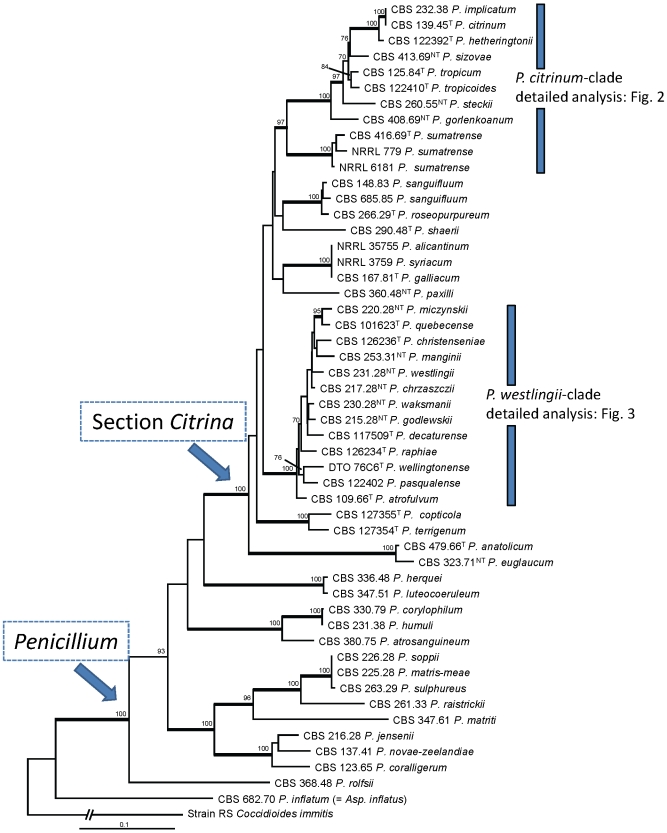
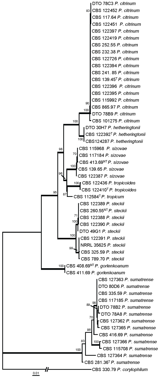
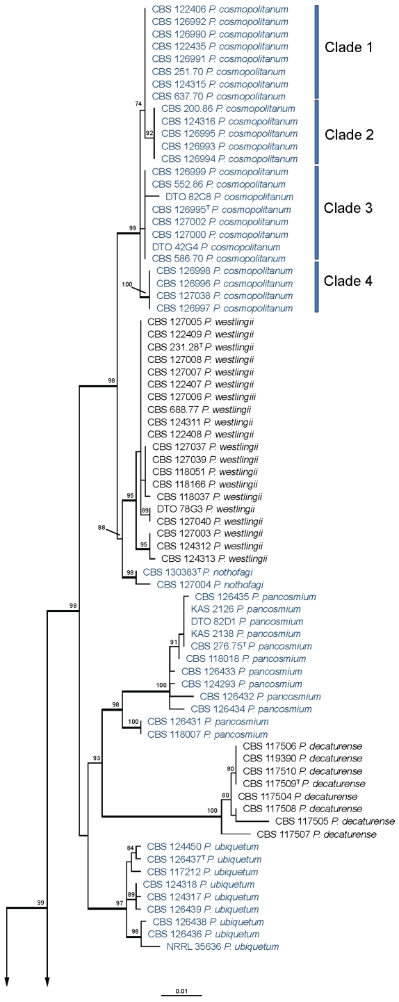
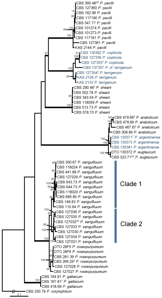
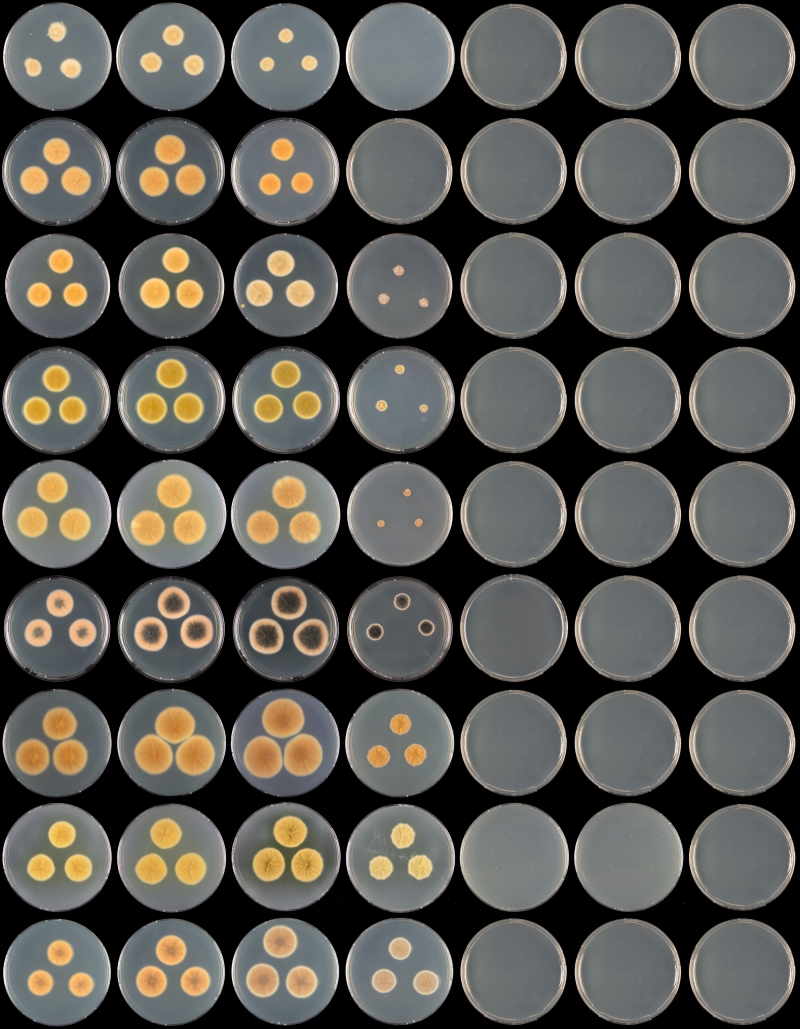
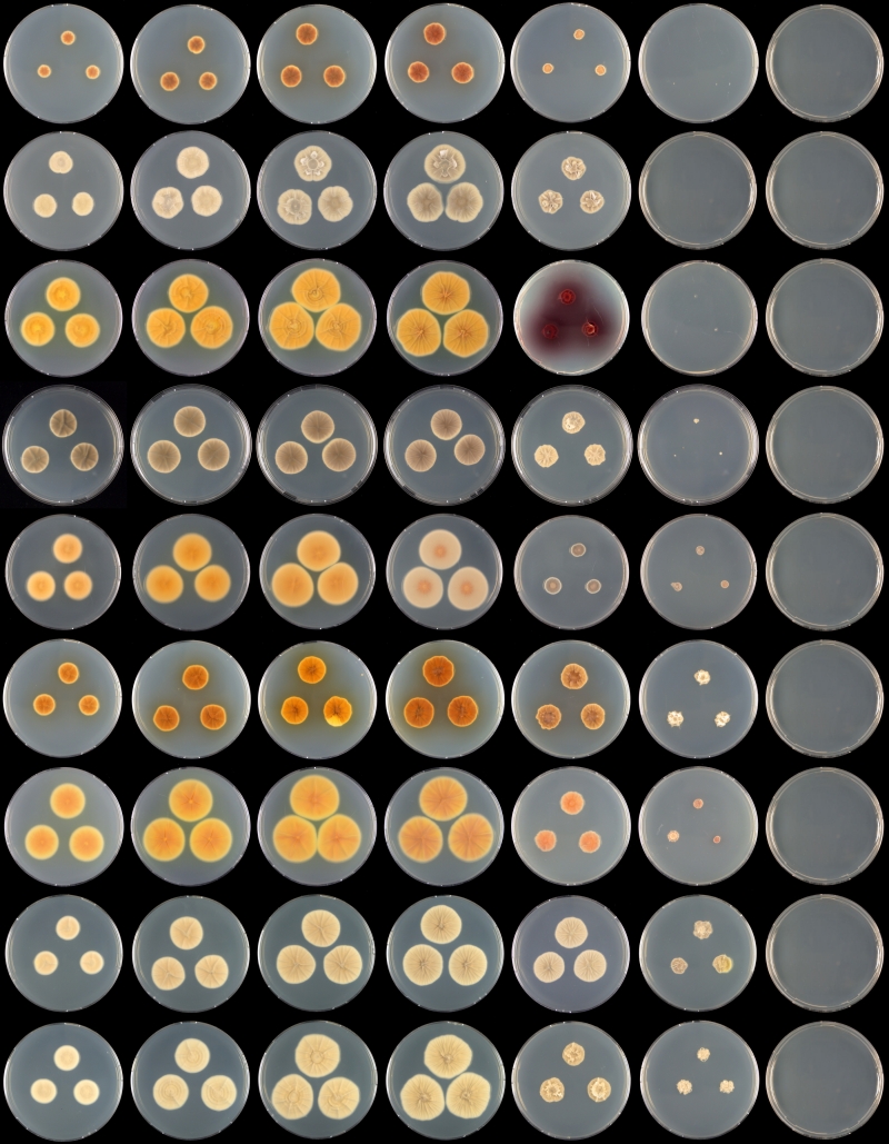
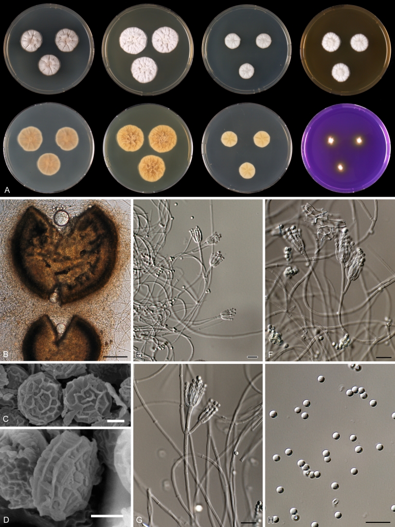
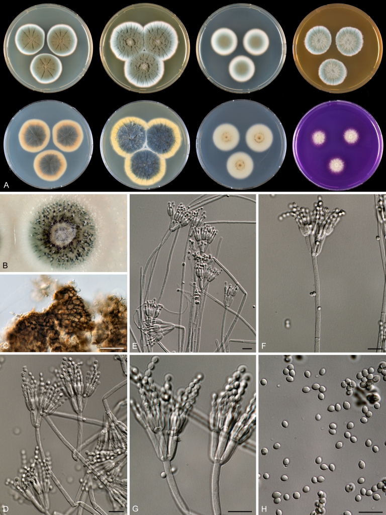
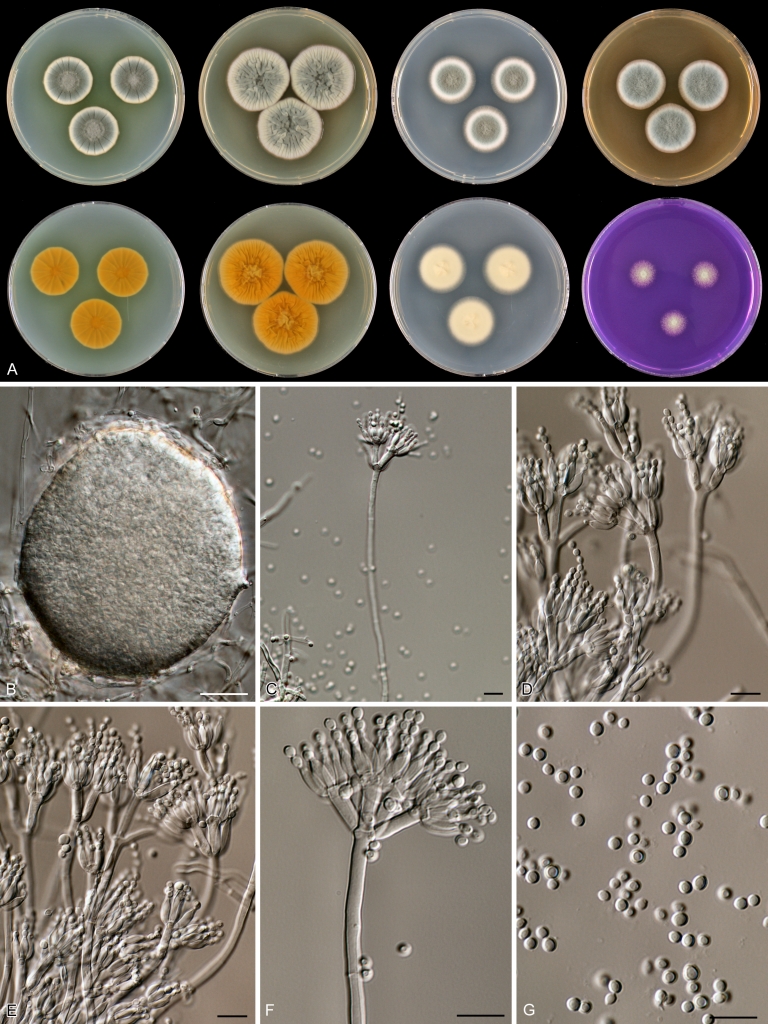
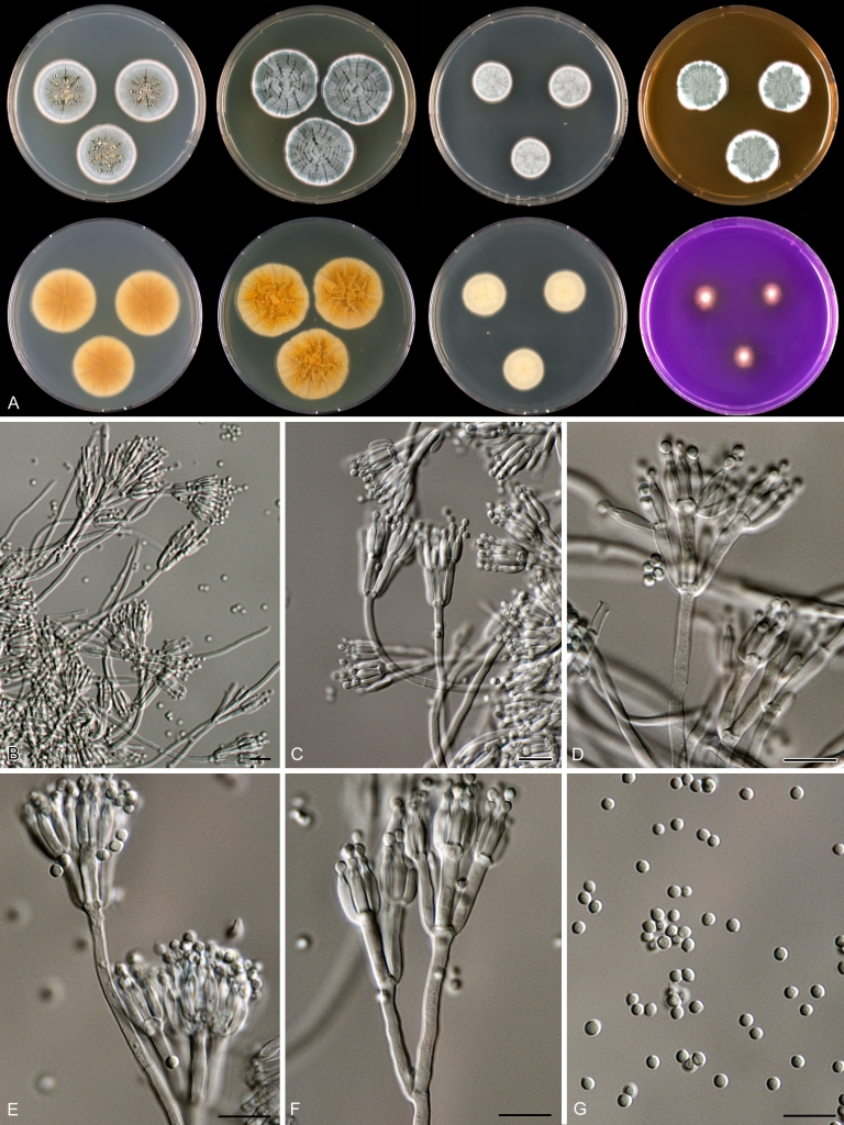
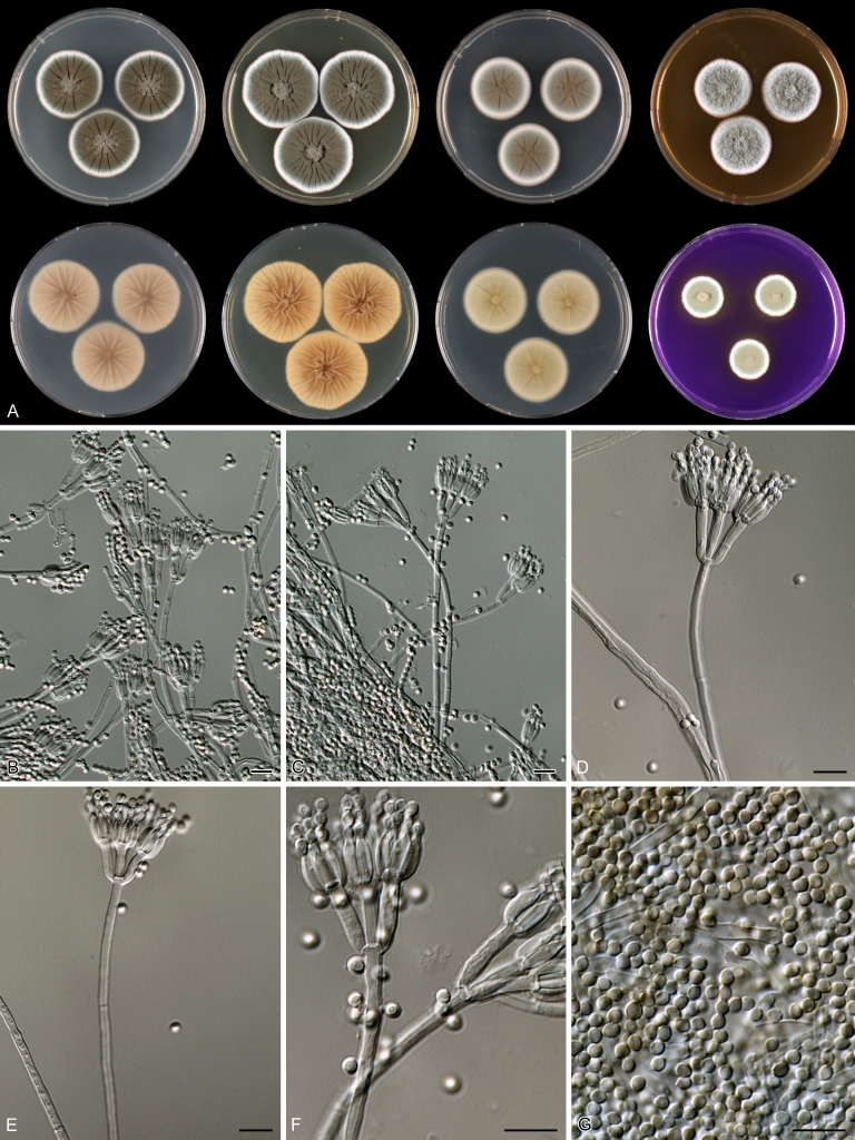
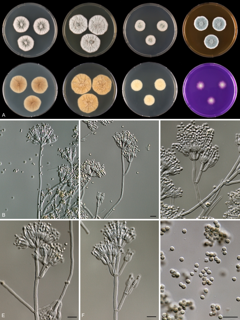
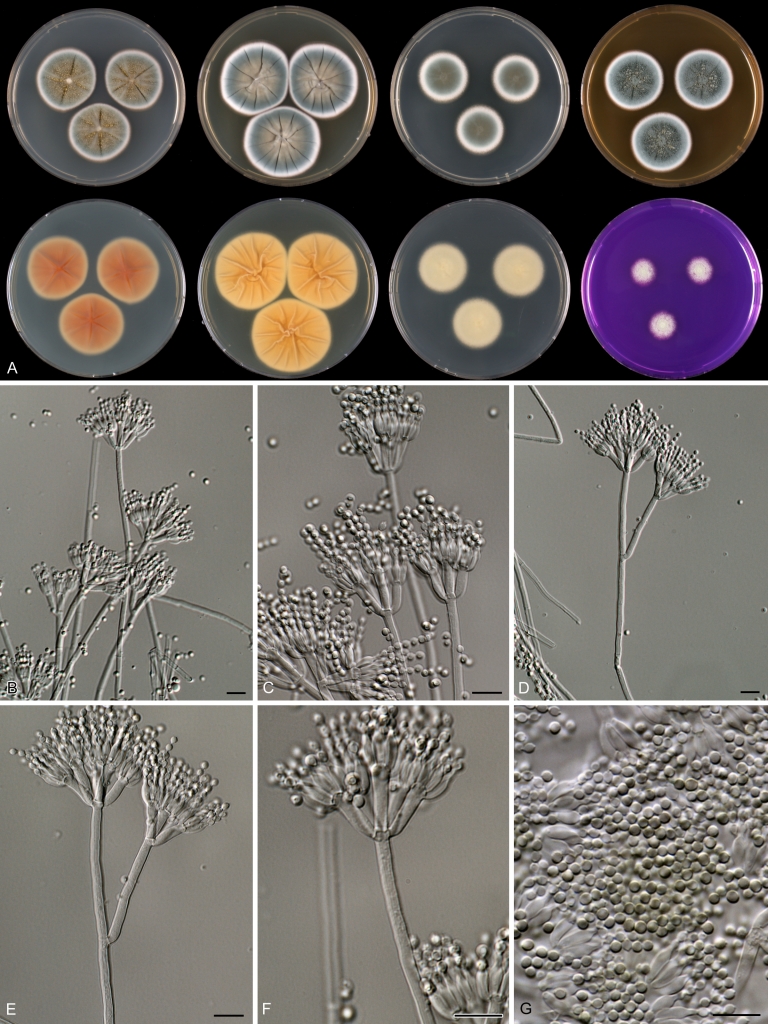
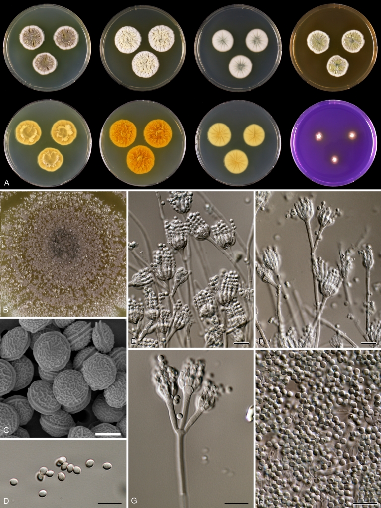
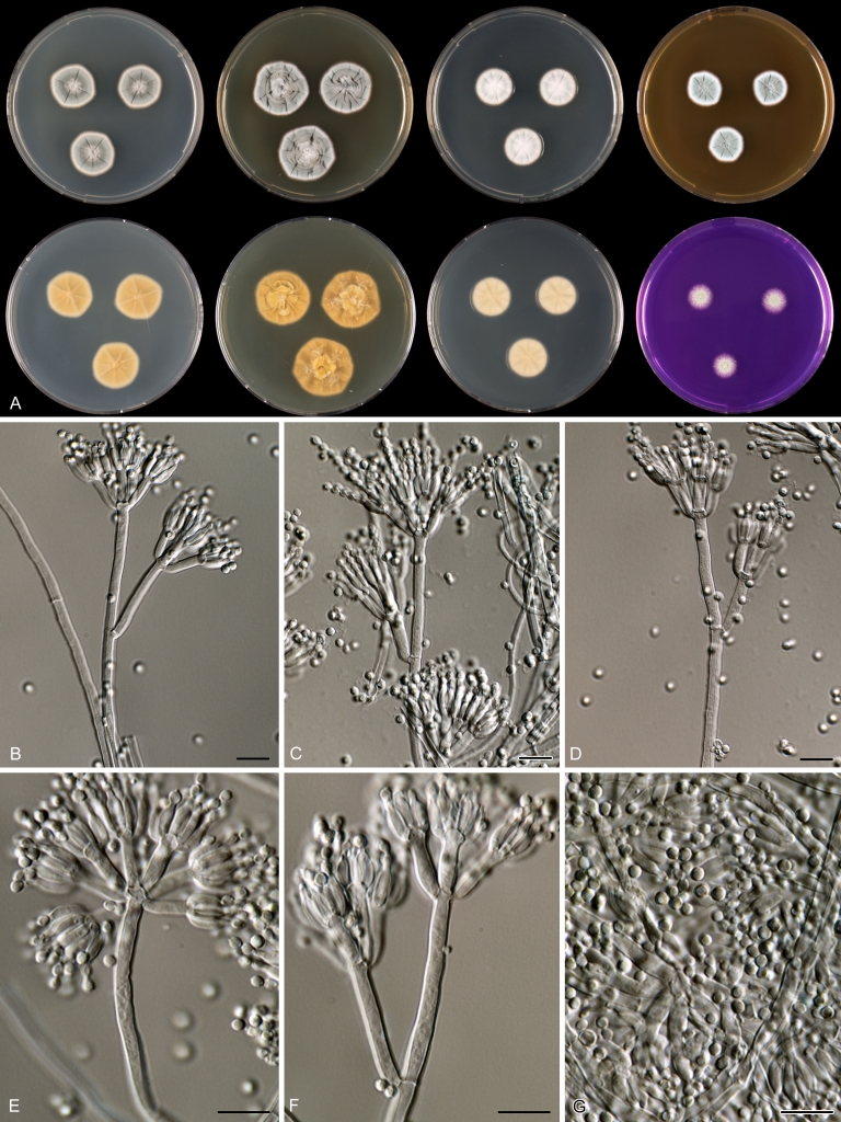
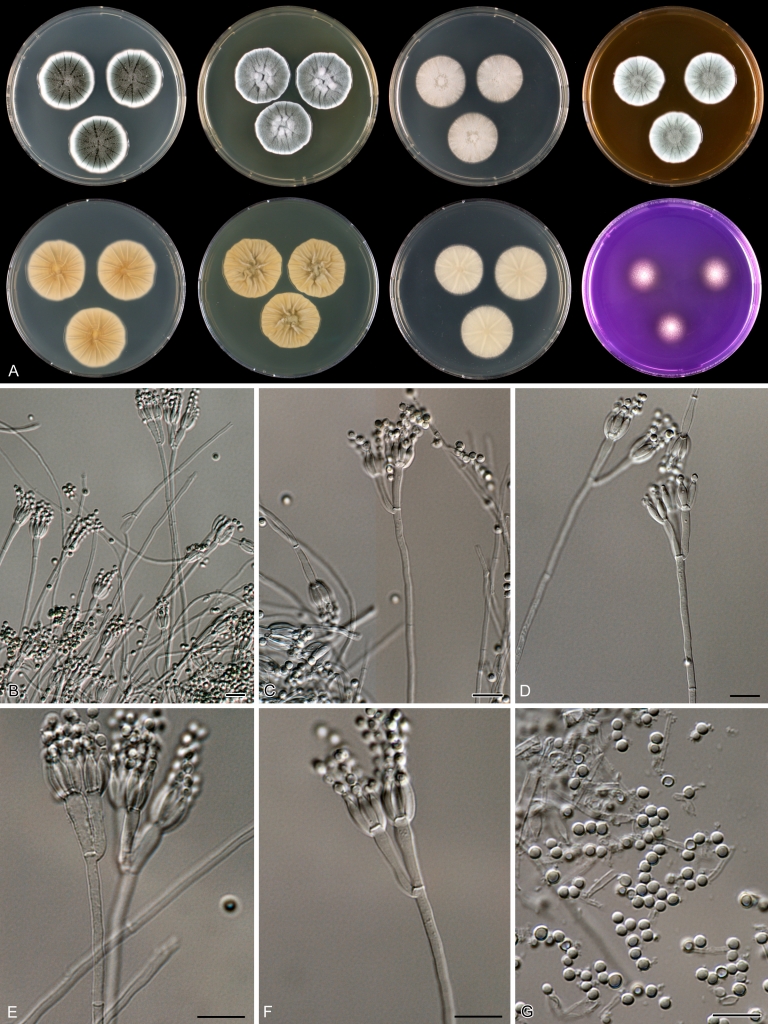
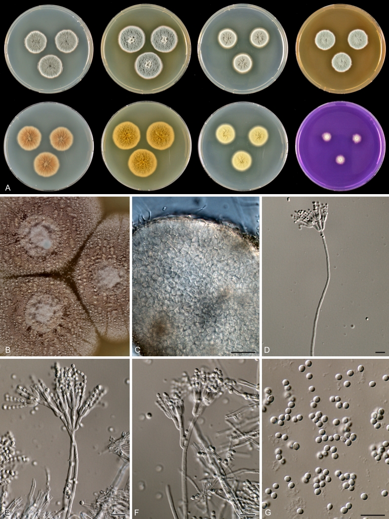
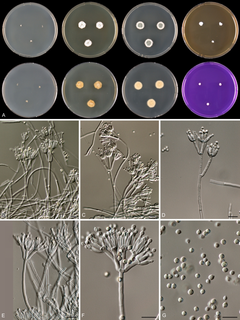
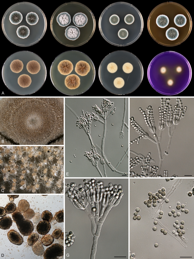
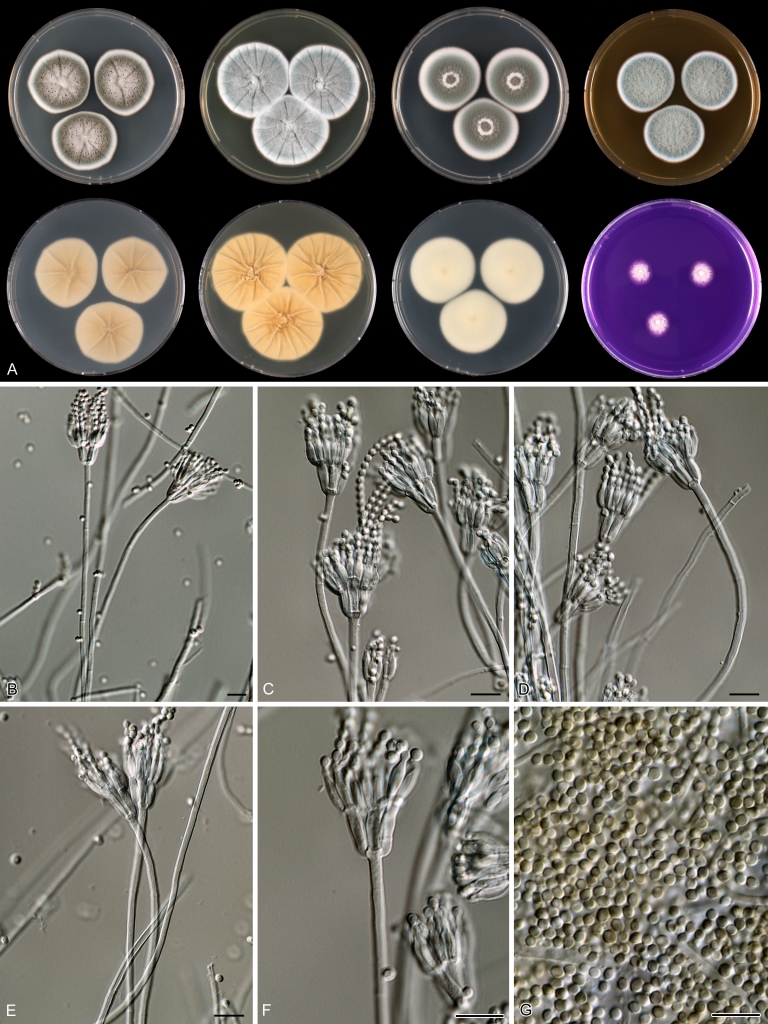
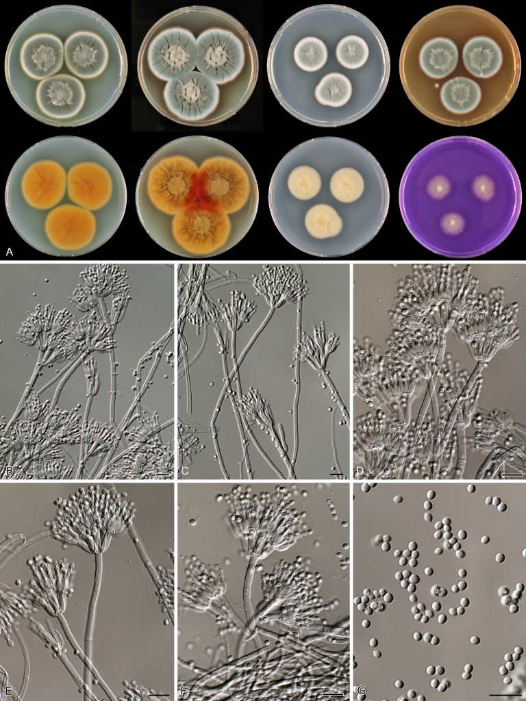
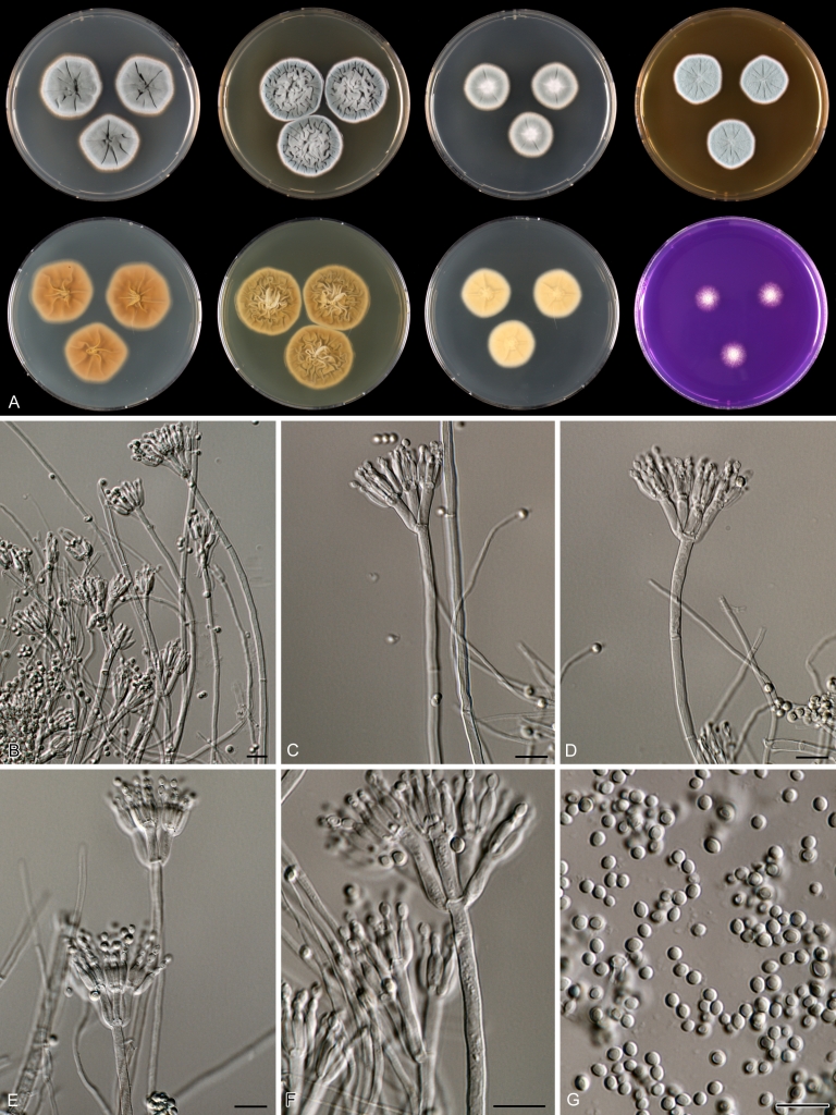
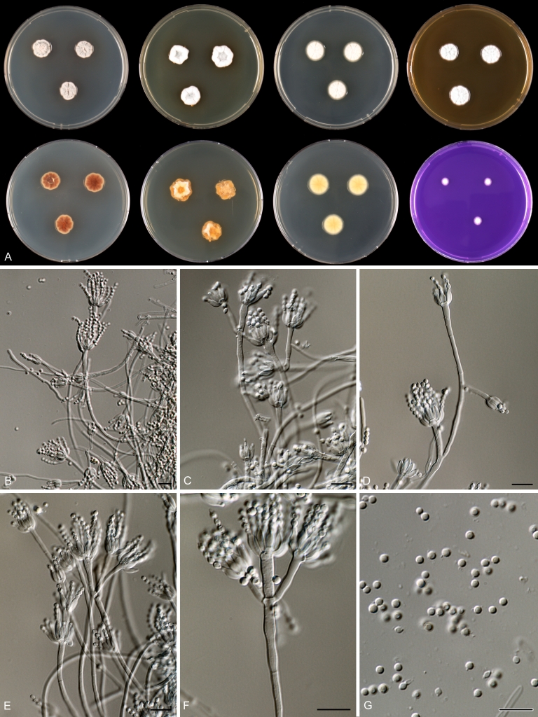
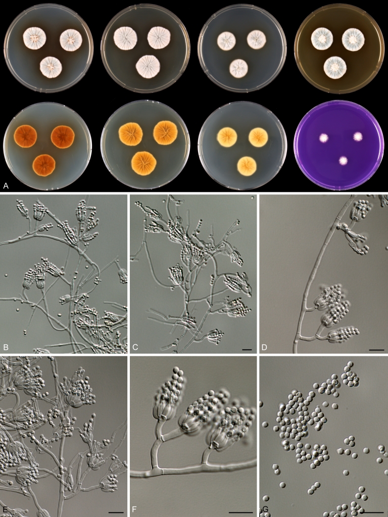
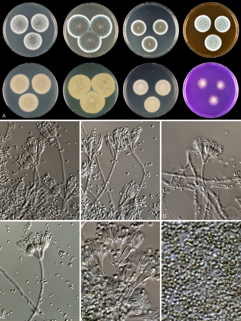
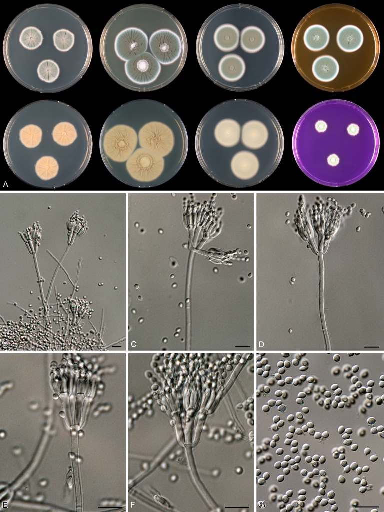
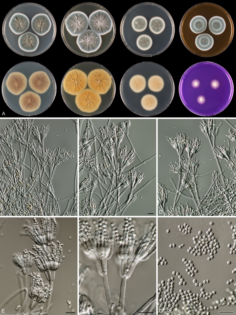
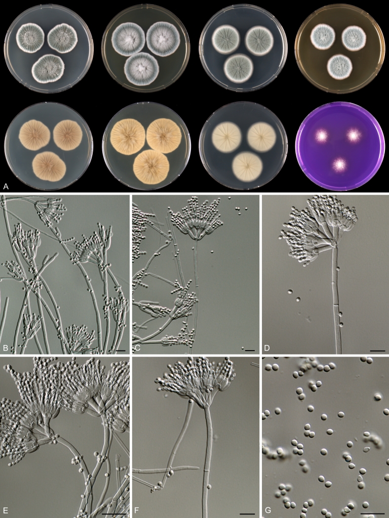
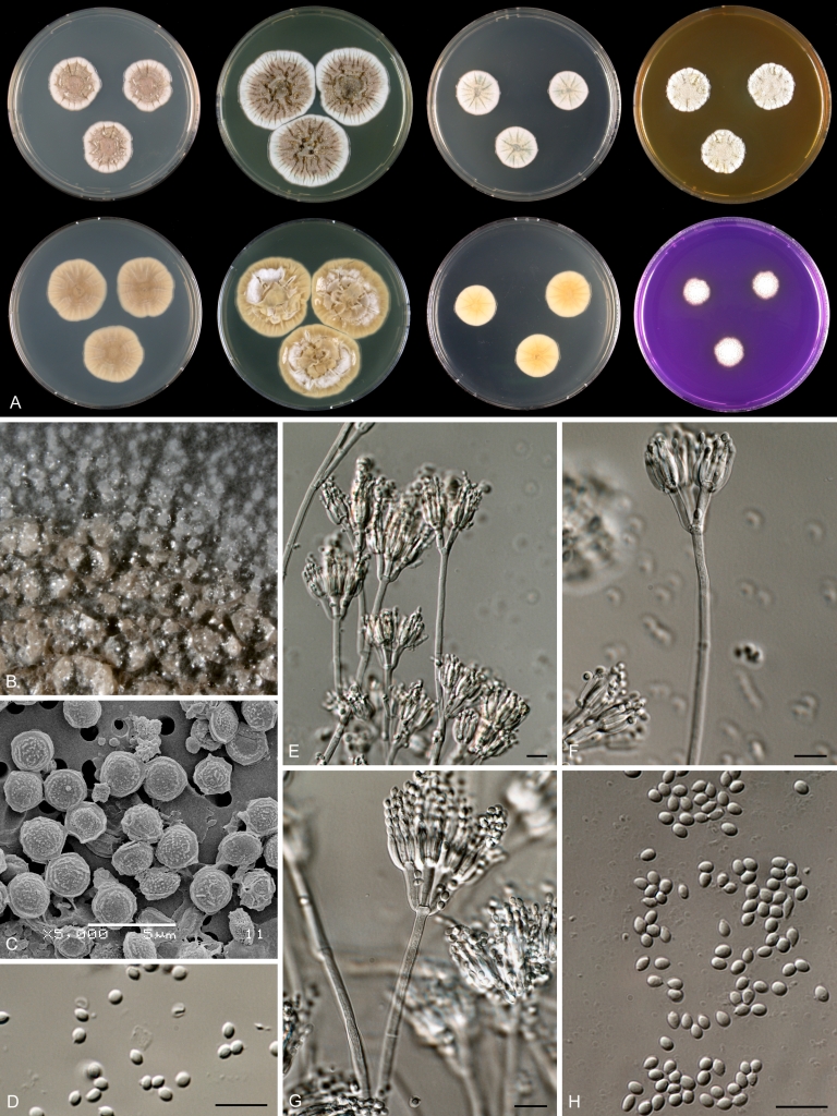
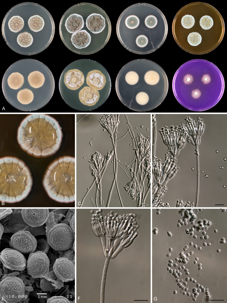
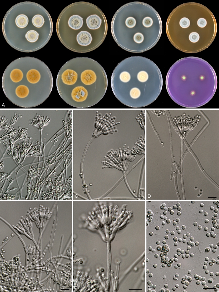
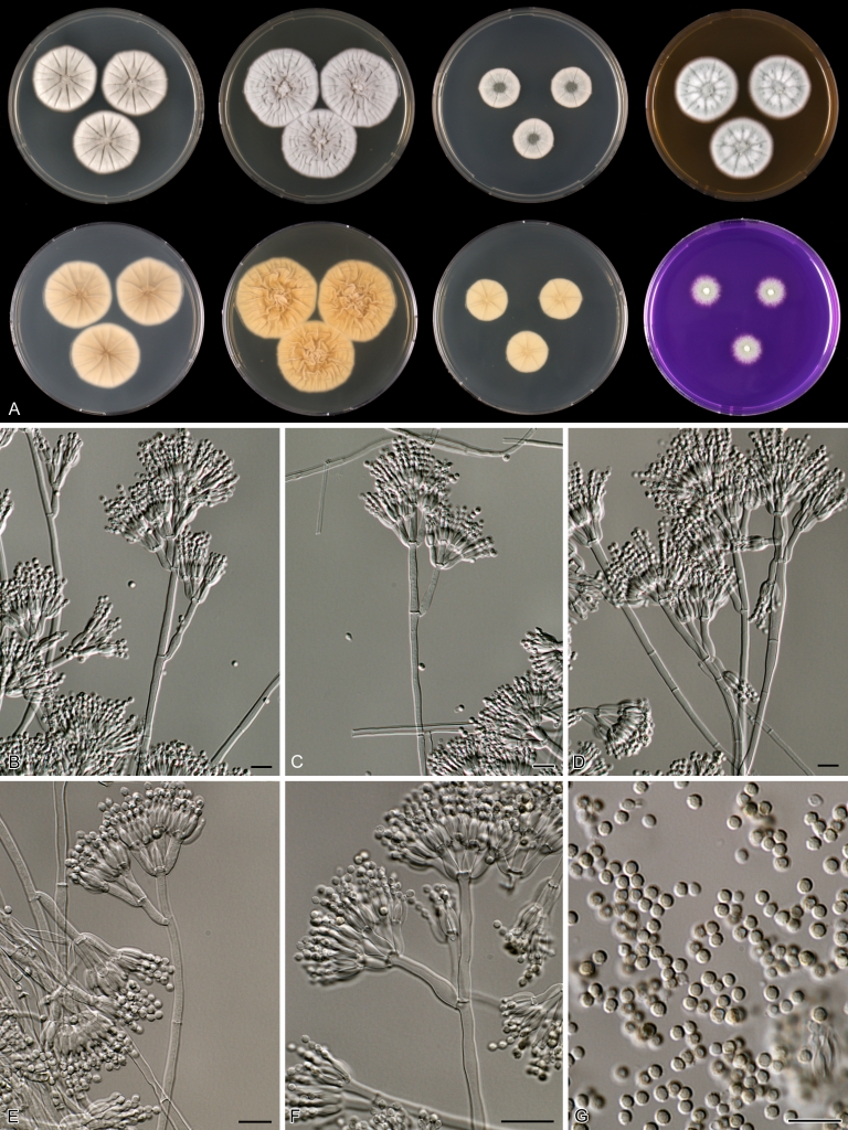
Species of Penicillium section Citrina have a worldwide distribution and occur commonly in soils. The section is here delimited using a combination of phenotypic characters and sequences of the nuclear ribosomal RNA gene operon, including the internal transcribed spacer regions ITS1 and ITS2, the 5.8S nrDNA (ITS) and partial RPB2 sequences. Species assigned to section Citrina share the production of symmetrically biverticillate conidiophores, flask shaped phialides (7.0-9.0 μm long) and relatively small conidia (2.0-3.0 μm diam). Some species can produce greyish-brown coloured cleistothecia containing flanged ascospores. In the present study, more than 250 isolates presumably belonging to section Citrina were examined using a combined analysis of phenotypic and physiological characters, extrolite profiles and ITS, β-tubulin and/or calmodulin sequences. Section Citrina includes 39 species, and 17 of those are described here as new. The most important phenotypic characters for distinguishing species are growth rates and colony reverse colours on the agar media CYA, MEA and YES; shape, size and ornamentation of conidia and the production of sclerotia or cleistothecia. Temperature-growth profiles were made for all examined species and are a valuable character characters for species identification. Species centered around P. citrinum generally have a higher maximum growth temperature (33-36 °C) than species related to P. westlingii (27-33 °C). Extrolite patterns and partial calmodulin and β-tubulin sequences can be used for sequence based identification and resolved all species. In contrast, ITS sequences were less variable and only 55 % of the species could be unambiguously identified with this locus.

Taxonomic novelties: Penicillium argentinense Houbraken, Frisvad & Samson, P. atrofulvum Houbraken, Frisvad & Samson, P. aurantiacobrunneum Houbraken, Frisvad & Samson, P. cairnsense Houbraken, Frisvad & Samson, P. christenseniae Houbraken, Frisvad & Samson, P. copticola Houbraken, Frisvad & Samson, P. cosmopolitanum Houbraken, Frisvad & Samson, P. neomiczynskii Cole, Houbraken, Frisvad & Samson, P. nothofagi Houbraken, Frisvad & Samson, P. pancosmium Houbraken, Frisvad & Samson, P. pasqualense Houbraken, Frisvad & Samson, P. quebecense Seifert, Houbraken, Frisvad & Samson, P. raphiae Houbraken, Frisvad & Samson, P. terrigenum Seifert, Houbraken, Frisvad & Samson, P. ubiquetum Houbraken, Frisvad & Samson, P. vancouverense Houbraken, Frisvad & Samson, P. wellingtonense Cole, Houbraken, Frisvad & Samson.

Keywords: citreoviridin; citrinin; phylogeny; soil fungi; taxonomy.


Figures

Fig. 1.
Fig. 1.
Best-scoring Maximum Likelihood tree using RAxML based on a combination of partial RPB2 and ITS sequences. Members of section Citrina are in a well-supported lineage (100 % bs) and some species previously belonging to series Citrina are placed in other lineages. Bootstrap percentages of the Maximum Likelihood (ML) analysis are presented at the nodes. Values less than 70 % supported in the ML are not shown and branches with more than 95 % bootstrap support are thickened. The bar indicates the number of substitutions per site. The phylogram is rooted with Coccidioides immitis (Strain RS).
Fig. 2.
Fig. 2.
Best-scoring Maximum Likelihood tree using RAxML based on a combination of partial β-tubulin and calmodulin sequences, showing the relationship among members of the P. citrinum-clade. Bootstrap percentages of the maximum likelihood (ML) analysis are presented at the nodes. Values less than 70 % supported in the ML are not shown and branches with more than 95 % bootstrap support are thickened. The bar indicates the number of substitutions per site. The phylogram is rooted with P. corylophilum (CBS 330.79).
Fig. 3.
Fig. 3.
Best-scoring Maximum Likelihood tree using RAxML based on a combination of partial β-tubulin and calmodulin sequences, showing the phylogenetic relationship among members of the P. westlingii-clade. Newly described species belonging to this section are presented in dark blue. Bootstrap percentages of the maximum likelihood (ML) analysis are presented at the nodes. Values less than 70 % supported in the ML are not shown and branches with more than 95 % bootstrap support are thickened. The bar indicates the number of substitutions per site. The phylogram is rooted with P. corylophilum (CBS 330.79).
Fig. 3.
Fig. 3.
Best-scoring Maximum Likelihood tree using RAxML based on a combination of partial β-tubulin and calmodulin sequences, showing the phylogenetic relationship among members of the P. westlingii-clade. Newly described species belonging to this section are presented in dark blue. Bootstrap percentages of the maximum likelihood (ML) analysis are presented at the nodes. Values less than 70 % supported in the ML are not shown and branches with more than 95 % bootstrap support are thickened. The bar indicates the number of substitutions per site. The phylogram is rooted with P. corylophilum (CBS 330.79).
Fig. 4.
Fig. 4.
Best-scoring Maximum Likelihood tree using RAxML based on a combination of partial β-tubulin and calmodulin sequences, showing the phylogenetic relationship among selected members of section Citrina. Newly described species belonging to this section are presented in dark blue. Bootstrap percentages of the maximum likelihood (ML) analysis are presented at the nodes. Values less than 70 % supported in the ML are not shown and branches with more than 95 % bootstrap support are thickened. The bar indicates the number of substitutions per site. The phylogram is rooted with P. corylophilum (CBS 330.79).
Fig. 5.
Fig. 5.
Overview of growth rates on CYA (reverse) after 7 d at various temperatures. Row, left to right: 21, 24, 27, 30, 33, 36 °C; columns, top to bottom: P. nothofagi, P. wellingtonense, P. godlewskii, P. vancouverense, P. neomiczynskii, P. atrofulvum, P. christenseniae, P. miczynskii, P. waksmanii.
Fig. 6.
Fig. 6.
Overview of growth rates on CYA (reverse) after 7 d at various temperatures. Row, left to right: 21, 24, 27, 30, 33, 36 °C; columns, top to bottom: P. raphiae, P. chrzaszczii, P. ubiquetum, P. aurantiacobrunneum, P. pancosmium, P. cosmopolitanum, P. westlingii, P. manginii, P. manginii.
Fig. 7.
Fig. 7.
Overview of growth rates on CYA (reverse) after 7 d at various temperatures. Row, left to right: 21, 24, 27, 30, 33, 36 °C; columns, top to bottom: P. roseopurpureum, P. tropicoides, P. cairnsense, P. pasqualense, P. decaturense, P. sanguifluum, P. quebecense, P. terrigenum, P. copticola.
Fig. 8.
Fig. 8.
Overview of growth rates on CYA (reverse) after 7 d at various temperatures. Row, left to right: 21, 24, 27, 30, 33, 36 °C; columns, top to bottom: P. paxilli, P. tropicum, P. sumatrense, P. gorlenkoanum, P. steckii, P. sizovae, P. argentinense, P. euglaucum, P. anatolicum.
Fig. 9.
Fig. 9.
Overview of growth rates on CYA (reverse) after 7 d at various temperatures. Row, left to right: 21, 24, 27, 30, 33, 36 °C; columns, top to bottom: P. gallaicum, P. hetheringtonii, P. citrinum, P. shearii.
Fig. 10.
Fig. 10.
Penicillium anatolicum. A. 7 d old cultures, 25 °C, left to right; first row, all obverse, CYA, YES, DG18, MEA; second row, CYA reverse, YES reverse, DG18 reverse, CREA obverse. B–C. Ascomata. D–E. Ascospores. F–H. Conidiophores. I. Conidia. Scale bars = 10 μm.
Fig. 11.
Fig. 11.
Penicillium argentinense. A. 7 d old cultures, 25 °C, left to right; first row, all obverse, CYA, YES, DG18, MEA; second row, CYA reverse, YES reverse, DG18 reverse, CREA obverse. B. Ascomata. C–D. Ascospores. E–G. Conidiophores. H. Conidia. Scale bars = 10 μm.
Fig. 12.
Fig. 12.
Penicillium atrofulvum. A. 7 d old cultures, 25 °C, left to right; first row, all obverse, CYA, YES, DG18, MEA; second row, CYA reverse, YES reverse, DG18 reverse, CREA obverse. B–C. Sclerotia. D–G. Conidiophores. H. Conidia. Scale bars = 10 μm.
Fig. 13.
Fig. 13.
Penicillium aurantiacobrunneum. A. 7 d old cultures, 25 °C, left to right; first row, all obverse, CYA, YES, DG18, MEA; second row, CYA reverse, YES reverse, DG18 reverse, CREA obverse. B. Sclerotia. C–F. Conidiophores. G. Conidia. Scale bars = 10 μm.
Fig. 14.
Fig. 14.
Penicillium cairnsense. A. 7 d old cultures, 25 °C, left to right; first row, all obverse, CYA, YES, DG18, MEA; second row, CYA reverse, YES reverse, DG18 reverse, CREA obverse. B–C. Sclerotia. D–G. Conidiophores. H. Conidia. Scale bars = 10 μm.
Fig. 15.
Fig. 15.
Penicillium christenseniae. A. 7 d old cultures, 25 °C, left to right; first row, all obverse, CYA, YES, DG18, MEA; second row, CYA reverse, YES reverse, DG18 reverse, CREA obverse. B–F. Conidiophores. G. Conidia. Scale bars = 10 μm.
Fig. 16.
Fig. 16.
Penicillium chrzaszczii. A. 7 d old cultures, 25 °C, left to right; first row, all obverse, CYA, YES, DG18, MEA; second row, CYA reverse, YES reverse, DG18 reverse, CREA obverse. B–F. Conidiophores. G. Conidia. Scale bars = 10 μm.
Fig. 17.
Fig. 17.
Penicillium citrinum. A. 7 d old cultures, 25 °C, left to right; first row, all obverse, CYA, YES, DG18, MEA; second row, CYA reverse, YES reverse, DG18 reverse, CREA obverse. B–F. Conidiophores. G. Conidia. Scale bars = 10 μm.
Fig. 18.
Fig. 18.
Penicillium copticola. A. 7 d old cultures, 25 °C, left to right; first row, all obverse, CYA, YES, DG18, MEA; second row, CYA reverse, YES reverse, DG18 reverse, CREA obverse. B–F. Conidiophores. G. Conidia. Scale bars = 10 μm.
Fig. 19.
Fig. 19.
Penicillium cosmopolitanum. A. 7 d old cultures, 25 °C, left to right; first row, all obverse, CYA, YES, DG18, MEA; second row, CYA reverse, YES reverse, DG18 reverse, CREA obverse. B–F. Conidiophores. G. Conidia. Scale bars = 10 μm.
Fig. 20.
Fig. 20.
Penicillium decaturense. A. 7 d old cultures, 25 °C, left to right; first row, all obverse, CYA, YES, DG18, MEA; second row, CYA reverse, YES reverse, DG18 reverse, CREA obverse. B–F. Conidiophores. G. Conidia. Scale bars = 10 μm.
Fig. 21.
Fig. 21.
Penicillium euglaucum. A. 7 d old cultures, 25 °C, left to right; first row, all obverse, CYA, YES, DG18, MEA; second row, CYA reverse, YES reverse, DG18 reverse, CREA obverse. B. Ascomata. C–D. Ascospores. E–G. Conidiophores. H. Conidia. Scale bars = 10 μm.
Fig. 22.
Fig. 22.
Penicillium gallaicum. A. 7 d old cultures, 25 °C, left to right; first row, all obverse, CYA, YES, DG18, MEA; second row, CYA reverse, YES reverse, DG18 reverse, CREA obverse. B. Sclerotia. C–F. Conidiophores. G. Conidia. Scale bars = 10 μm.
Fig. 23.
Fig. 23.
Penicillium godlewskii. A. 7 d old cultures, 25 °C, left to right; first row, all obverse, CYA, YES, DG18, MEA; second row, CYA reverse, YES reverse, DG18 reverse, CREA obverse. B–F. Conidiophores. G. Conidia. Scale bars = 10 μm.
Fig. 24.
Fig. 24.
Penicillium gorlenkoanum. A. 7 d old cultures, 25 °C, left to right; first row, all obverse, CYA, YES, DG18, MEA; second row, CYA reverse, YES reverse, DG18 reverse, CREA obverse. B–F. Conidiophores. G. Conidia. Scale bars = 10 μm.
Fig. 25.
Fig. 25.
Penicillium hetheringtonii. A. 7 d old cultures, 25 °C, left to right; first row, all obverse, CYA, YES, DG18, MEA; second row, CYA reverse, YES reverse, DG18 reverse, CREA obverse. B–F. Conidiophores. G. Conidia. Scale bars = 10 μm.
Fig. 26.
Fig. 26.
Penicillium manginii. A. 7 d old cultures, 25 °C, left to right; first row, all obverse, CYA, YES, DG18, MEA; second row, CYA reverse, YES reverse, DG18 reverse, CREA obverse. B–C. Sclerotia. D–G. Conidiophores. H. Conidia. Scale bars = 10 μm.
Fig. 27.
Fig. 27.
Penicillium miczynskii. A. 7 d old cultures, 25 °C, left to right; first row, all obverse, CYA, YES, DG18, MEA; second row, CYA reverse, YES reverse, DG18 reverse, CREA obverse. B–C. Sclerotia. D–F. Conidiophores. G. Conidia. Scale bars = 10 μm.
Fig. 28.
Fig. 28.
Penicillium neomiczynskii. A. 7 d old cultures, 25 °C, left to right; first row, all obverse, CYA, YES, DG18, MEA; second row, CYA reverse, YES reverse, DG18 reverse, CREA obverse. B–F. Conidiophores. G. Conidia. Scale bars = 10 μm.
Fig. 29.
Fig. 29.
Penicillium nothofagi. A. 7 d old cultures, 25 °C, left to right; first row, all obverse, CYA, YES, DG18, MEA; second row, CYA reverse, YES reverse, DG18 reverse, CREA obverse. B–F. Conidiophores. G. Conidia. Scale bars = 10 μm.
Fig. 30.
Fig. 30.
Penicillium pancosmium. A. 7 d old cultures, 25 °C, left to right; first row, all obverse, CYA, YES, DG18, MEA; second row, CYA reverse, YES reverse, DG18 reverse, CREA obverse. B–F. Conidiophores. G. Conidia. Scale bars = 10 μm.
Fig. 31.
Fig. 31.
Penicillium pasqualense. A. 7 d old cultures, 25 °C, left to right; first row, all obverse, CYA, YES, DG18, MEA; second row, CYA reverse, YES reverse, DG18 reverse, CREA obverse. B–D. Sclerotia. E–G. Conidiophores. H. Conidia. Scale bars = 10 μm.
Fig. 32.
Fig. 32.
Penicillium paxilli. A. 7 d old cultures, 25 °C, left to right; first row, all obverse, CYA, YES, DG18, MEA; second row, CYA reverse, YES reverse, DG18 reverse, CREA obverse. B–F. Conidiophores. G. Conidia. Scale bars = 10 μm.
Fig. 33.
Fig. 33.
Penicillium quebecense. A. 7 d old cultures, 25 °C, left to right; first row, all obverse, CYA, YES, DG18, MEA; second row, CYA reverse, YES reverse, DG18 reverse, CREA obverse. B–F. Conidiophores. G. Conidia. Scale bars = 10 μm.
Fig. 34.
Fig. 34.
Penicillium raphiae. A. 7 d old cultures, 25 °C, left to right; first row, all obverse, CYA, YES, DG18, MEA; second row, CYA reverse, YES reverse, DG18 reverse, CREA obverse. B–F. Conidiophores. G. Conidia. Scale bars = 10 μm.
Fig. 35.
Fig. 35.
Penicillium roseopurpureum. A. 7 d old cultures, 25 °C, left to right; first row, all obverse, CYA, YES, DG18, MEA; second row, CYA reverse, YES reverse, DG18 reverse, CREA obverse. B–F. Conidiophores. G. Conidia. Scale bars = 10 μm.
Fig. 36.
Fig. 36.
Penicillium sanguifluum. A. 7 d old cultures, 25 °C, left to right; first row, all obverse, CYA, YES, DG18, MEA; second row, CYA reverse, YES reverse, DG18 reverse, CREA obverse. B–F. Conidiophores. G. Conidia. Scale bars = 10 μm.
Fig. 37.
Fig. 37.
Penicillium shearii. A. 7 d old cultures, 25 °C, left to right; first row, all obverse, CYA, YES, DG18, MEA; second row, CYA reverse, YES reverse, DG18 reverse, CREA obverse. B. Sclerotia. C–D. Ascospores. E–G. Conidiophores. H. Conidia. Scale bars = 10 μm.
Fig. 38.
Fig. 38.
Penicillium sizovae. A. 7 d old cultures, 25 °C, left to right; first row, all obverse, CYA, YES, DG18, MEA; second row, CYA reverse, YES reverse, DG18 reverse, CREA obverse. B–F. Conidiophores. G. Conidia. Scale bars = 10 μm.
Fig. 39.
Fig. 39.
Penicillium steckii. A. 7 d old cultures, 25 °C, left to right; first row, all obverse, CYA, YES, DG18, MEA; second row, CYA reverse, YES reverse, DG18 reverse, CREA obverse. B–F. Conidiophores. G. Conidia. Scale bars = 10 μm.
Fig. 40.
Fig. 40.
Penicillium sumatrense. A. 7 d old cultures, 25 °C, left to right; first row, all obverse, CYA, YES, DG18, MEA; second row, CYA reverse, YES reverse, DG18 reverse, CREA obverse. B–F. Conidiophores. G. Conidia. Scale bars = 10 μm.
Fig. 41.
Fig. 41.
Penicillium terrigenum. A. 7 d old cultures, 25 °C, left to right; first row, all obverse, CYA, YES, DG18, MEA; second row, CYA reverse, YES reverse, DG18 reverse, CREA obverse. B–F. Conidiophores. G. Conidia. Scale bars = 10 μm.
Fig. 42.
Fig. 42.
Penicillium tropicoides. A. 7 d old cultures, 25 °C, left to right; first row, all obverse, CYA, YES, DG18, MEA; second row, CYA reverse, YES reverse, DG18 reverse, CREA obverse. B. Ascomata. C–D. Ascospores. E–G. Conidiophores. H. Conidia. Scale bars = 10 μm.
Fig. 43.
Fig. 43.
Penicillium tropicum. A. 7 d old cultures, 25 °C, left to right; first row, all obverse, CYA, YES, DG18, MEA; second row, CYA reverse, YES reverse, DG18 reverse, CREA obverse. B. Sclerotia. C. Ascospores. D–F. Conidiophores. G. Conidia. Scale bars = 10 μm.
Fig. 44.
Fig. 44.
Penicillium ubiquetum. A. 7 d old cultures, 25 °C, left to right; first row, all obverse, CYA, YES, DG18, MEA; second row, CYA reverse, YES reverse, DG18 reverse, CREA obverse. B–F. Conidiophores. G. Conidia. Scale bars = 10 μm.
Fig. 45.
Fig. 45.
Penicillium vancouverense. A. 7 d old cultures, 25 °C, left to right; first row, all obverse, CYA, YES, DG18, MEA; second row, CYA reverse, YES reverse, DG18 reverse, CREA obverse. B–F. Conidiophores. G. Conidia. Scale bars = 10 μm.
Fig. 46.
Fig. 46.
Penicillium waksmanii. A. 7 d old cultures, 25 °C, left to right; first row, all obverse, CYA, YES, DG18, MEA; second row, CYA reverse, YES reverse, DG18 reverse, CREA obverse. B–F. Conidiophores. G. Conidia. Scale bars = 10 μm.
Fig. 47.
Fig. 47.
Penicillium wellingtonense. A. 7 d old cultures, 25 °C, left to right; first row, all obverse, CYA, YES, DG18, MEA; second row, CYA reverse, YES reverse, DG18 reverse, CREA obverse. B–F. Conidiophores. G. Conidia. Scale bars = 10 μm.
Fig. 48.
Fig. 48.
Penicillium westlingii. A. 7 d old cultures, 25 °C, left to right; first row, all obverse, CYA, YES, DG18, MEA; second row, CYA reverse, YES reverse, DG18 reverse, CREA obverse. B–F. Conidiophores. G. Conidia. Scale bars = 10 μm.

References

    1. Abe S. (1956). Studies on the classification of the Penicillia. Journal of General and Applied Microbiology 2: 1–193
    1. Baghdadi VC. (1968). De speciebus novis Penicilli Fr. et Aspergilli Fr. E terries Syriae isolatis notula. Novitates Systematicae Plantarum non Vascularium 7: 96–114
    1. Belofsky GN, Gloer JB, Wicklow DT, Dowd PF. (1995). Anti-insectan alkaloids. Shearinines A-C and a new paxilline derivative from the ascostromata of Eupenicillium shearii. Tetrahedron 51: 3959–3968
    1. Beyma Thoe Kingma FH van. (1940). Beschreibung einiger neuer Pilzarten aus dem Centraalbureau voor Schimmelcultures, Baarn (Nederland) VI. Antonie van Leeuwenhoek 6: 263–290 - PubMed
    1. Brefeld O. (1874). Botanische Untersuchungen über Schimmelpilze. Heft 2. “Die Entwicklungsgeschichte von Penicillium”. Leipzig: A. Felix;

LinkOut - more resources