. 2012 Jan;4(1):60-73.
doi: 10.18632/aging.100428.

Reprogrammed keratinocytes from elderly type 2 diabetes patients suppress senescence genes to acquire induced pluripotency

Affiliations

Reprogrammed keratinocytes from elderly type 2 diabetes patients suppress senescence genes to acquire induced pluripotency

Seiga Ohmine et al. Aging (Albany NY). 2012 Jan.

Abstract

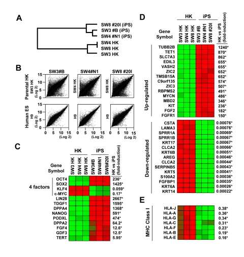
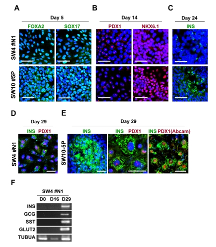
Nuclear reprogramming enables patient-specific derivation of induced pluripotent stem (iPS) cells from adult tissue. Yet, iPS generation from patients with type 2 diabetes (T2D) has not been demonstrated. Here, we report reproducible iPS derivation of epidermal keratinocytes (HK) from elderly T2D patients. Transduced with human OCT4, SOX2, KLF4 and c-MYC stemness factors under serum-free and feeder-free conditions, reprogrammed cells underwent dedifferentiation with mitochondrial restructuring, induction of endogenous pluripotency genes - including NANOG, LIN28, and TERT, and down-regulation of cytoskeletal, MHC class I- and apoptosis-related genes. Notably, derived iPS clones acquired a rejuvenated state, characterized by elongated telomeres and suppressed senescence-related p15INK4b/p16INK4a gene expression and oxidative stress signaling. Stepwise guidance with lineage-specifying factors, including Indolactam V and GLP-1, redifferentiated HK-derived iPS clones into insulin-producing islet-like progeny. Thus, in elderly T2D patients, reprogramming of keratinocytes ensures a senescence-privileged status yielding iPS cells proficient for regenerative applications.


Conflict of interest statement

The authors of this manuscript have no conflict of interest to declare.

Figures

Figure 1
Figure 1. Expression of pluripotency-associated markers in HK-derived iPS clones
(A) Early-passage HK cells (left panel) were infected with lentivirus (LV) vector encoding OCT4, SOX2, KLF4 and c-MYC. Seven days post-infection (center panel), early iPS-like colonies were detected (right panel in higher magnification). (B) HK-derived iPS clones were either derived from patients who were non-diabetic (ND) or type 2 diabetic (T2D). iPS clones, cultured under feeder-free conditions, exhibited human ES-like morphologies, while expressing high levels of alkaline phosphatase (AP). (C) Patient HK-derived iPS clones were further characterized through immunocytochemistry analysis using a panel of pluripotency markers. All clones were negative for SSEA-1 expression, while staining positive for pluripotency markers SSEA-4, TRA-1-60, TRA-1-81, OCT4, SOX2, KLF4 and NANOG. Scale bars represent 100 μm.
Figure 2
Figure 2. Pluripotency of HK-derived iPS cells verified through spontaneous differentiation in vitro and in vivo
(A) HK-derived iPS clones were analyzed via immunocytochemistry for lineage markers for three germ layers (endoderm, mesoderm and ectoderm). Scale bars indicate 50 μm. (B) Transplant of HK-derived iPS cells into the kidney capsule of SCID-beige mice resulted in teratoma formation. Pictures of harvested kidneys (with or without iPS transplant) are shown. (C) H&E staining demonstrated multiple lineages within the complex architecture of the tumor, including muscle, adipose, immature neuroepithelium and glandular tissues.
Figure 3
Figure 3. Variations in gene expression profile upon induced pluripotency
(A) Dendrogram describing the unsupervised hierarchal clustering of patient-derived HK cells and HK-derived iPS cells. (B) Global gene expression patterns of HK-derived iPS clones were compared with their parental HK cells (upper panels), or with that of human embryonic stem cells (H9, lower panels, GSM190779), upon RNA microarray analysis. (C) Heatmap showing the up-regulation (red) and down-regulation (green) of pluripotency-associated genes in HK- and HK-derived iPS clones. The four factors used to induce pluripotency are indicated. The changes in gene expression levels in iPS cells, relative to those in parental HK cells, were calculated using microarray data from three parental HK cells and three HK-derived iPS cells, and shown as fold-induction in iPS cells. Statistically significant changes are indicated by asterisks (p<0.05). Notably, HK cells originally expressed high levels of endogenous KLF4 and c-MYC, resulting in down-regulation of these two key reprogramming factors in derived iPS cells. (D) Heatmap showing the top 15 genes which were up-regulated (upper panel) or down-regulated (lower panel) upon reprogramming. Statistically significant changes are indicated by asterisks (p<0.05). (E) Comparison of the major histocompatibility complex (MHC) class I gene expression profiles between HK and iPS cells. Statistically significant changes are indicated by asterisks (p<0.05).
Figure 4
Figure 4. Morphological variations of patient-derived iPS cells upon reprogramming
(A) High-resolution electron micrographs of HK cells before (SW4 parental HK and SW8 parental HK) and after (SW4 #N1, SW3 #B, SW8 #20I and SW10 #5P) induced pluripotency. Representative micrograph of a verified fibroblast-derived iPS cell is also included. Scale bars represent 2 μm. (B) Mitotic events of two iPS clones were shown (left panel in metaphase; right panel in anaphase). Scale bars represent 2 μm. (C) Endoplasmic reticulum and the Golgi structures in HK and HK-derived iPS cells are shown. Scale bars represent 0.5 μm. (D) Mature mitochondria with well-developed cristae in parental HK cells (SW8 parental) and immature mitochondria in iPS clones (SW3 #B, SW8 #20I and SW10 #5P) are indicated by arrows. Keratin intermediate filaments in parental HK cells are indicated by arrowheads. Scale bars represent 0.5 μm.
Figure 5
Figure 5. Comparison of telomerase activity, cellular senescence and programmed cell death in HK cells before and after induced pluripotency
(A) RT-PCR analysis of TERT-specific transcripts in parental HK cells and iPS clones. GAPDH was used as control. (B) Telomere lengths in HK and HK-derived iPS cells were determined by the terminal restriction fragment lengths. Southern blot analysis and corresponding telomere fragment lengths derived from densitometric quantification are shown. (C) Schematic representation of key senescence- and apoptosis-regulating pathways. (D) Changes in expression levels of key genes, involved in cellular senescence or apoptosis, were determined using the microarray data of three parental HK cells and three HK-derived iPS cells, and fold induction of individual genes in iPS cells, relative to those in parental HK cells, are shown. Statistically significant changes are indicated by asterisks (p<0.05).
Figure 6
Figure 6. Guided in vitro differentiation of patient iPS cells into insulin-producing islet-like cells
iPS cells, differentiated through step-wise differentiation, were analyzed by immunocytochemistry for stage-specific markers at day 5 (A), 14 (B), 24 (C) and 29 (D and E). Scale bars indicate 50 μm for A, B, C and E (left panel), 10 μm for D and E (middle and right panels) and an alternative antibody (Abcam, #ab47267) against PDX1 is shown in E (right panel). (F) RT-PCR analysis of the mRNA of SW4#N1 clone, harvested at differentiation day 0, 16 and 29, confirmed the expression of insulin (INS), glucagon (GCG), somatostatin (SST), glucose transporter 2 (GLUT2) on day 29. α-tubulin was used as control (TUBUA).

References

    1. Cowie CC, Rust KF, Ford ES, Eberhardt MS, Byrd-Holt DD, Li C, Williams DE, Gregg EW, Bainbridge KE, Saydah SH, Geiss LS. Full accounting of diabetes and pre-diabetes in the U.S. population in 1988-1994 and 2005-2006. Diabetes Care. 2009;32:287–294. - PMC - PubMed
    1. Terzic A, Waldman S. Chronic diseases: the emerging pandemic. Clin Transl Sci. 2011;4:225–226. - PMC - PubMed
    1. Kroon E, Martinson LA, Kadoya K, Bang AG, Kelly OG, Eliazer S, Young H, Richardson M, Smart NG, Cunningham J, Agulnick AD, D'Amour KA, Carpenter MK, Baetge EE. Pancreatic endoderm derived from human embryonic stem cells generates glucose-responsive insulin-secreting cells in vivo. Nat Biotechnol. 2008;26:443–452. - PubMed
    1. D'Amour KA, Bang AG, Eliazer S, Kelly OG, Agulnick AD, Smart NG, Moorman MA, Kroon E, Carpenter MK, Baetge EE. Production of pancreatic hormone-expressing endocrine cells from human embryonic stem cells. Nat Biotechnol. 2006;24:1392–1401. - PubMed
    1. Jiang J, Au M, Lu K, Eshpeter A, Korbutt G, Fisk G, Majumdar AS. Generation of insulin-producing islet-like clusters from human embryonic stem cells. Stem Cells. 2007;25:1940–1953. - PubMed

Publication types