. 2012 Mar 15;188(6):2516-23.
doi: 10.4049/jimmunol.1102213. Epub 2012 Feb 3.

Derivation and maintenance of virtual memory CD8 T cells

Affiliations

Derivation and maintenance of virtual memory CD8 T cells

Adovi D Akue et al. J Immunol. .

Erratum in

  • J Immunol. 2014 Sep 1;193(5):2609

Abstract

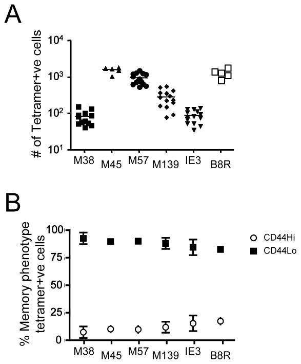
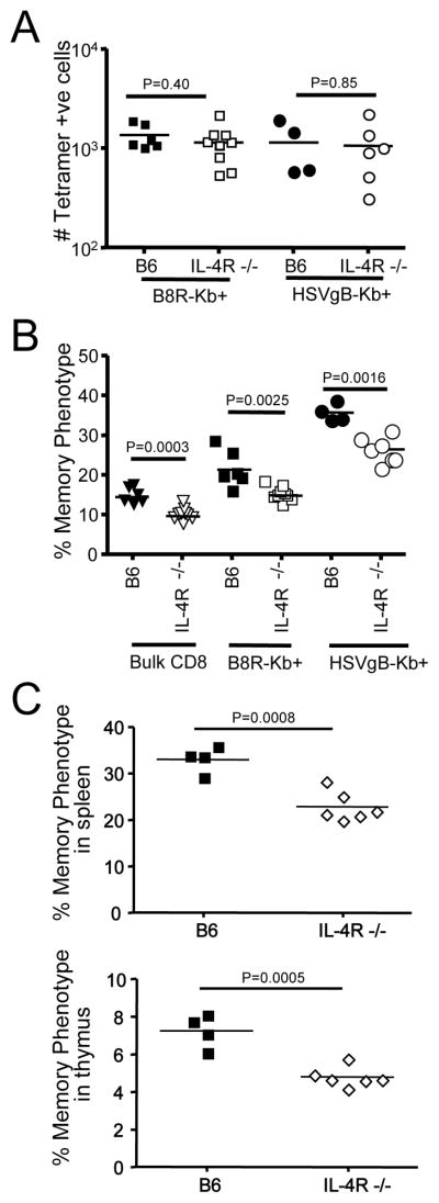
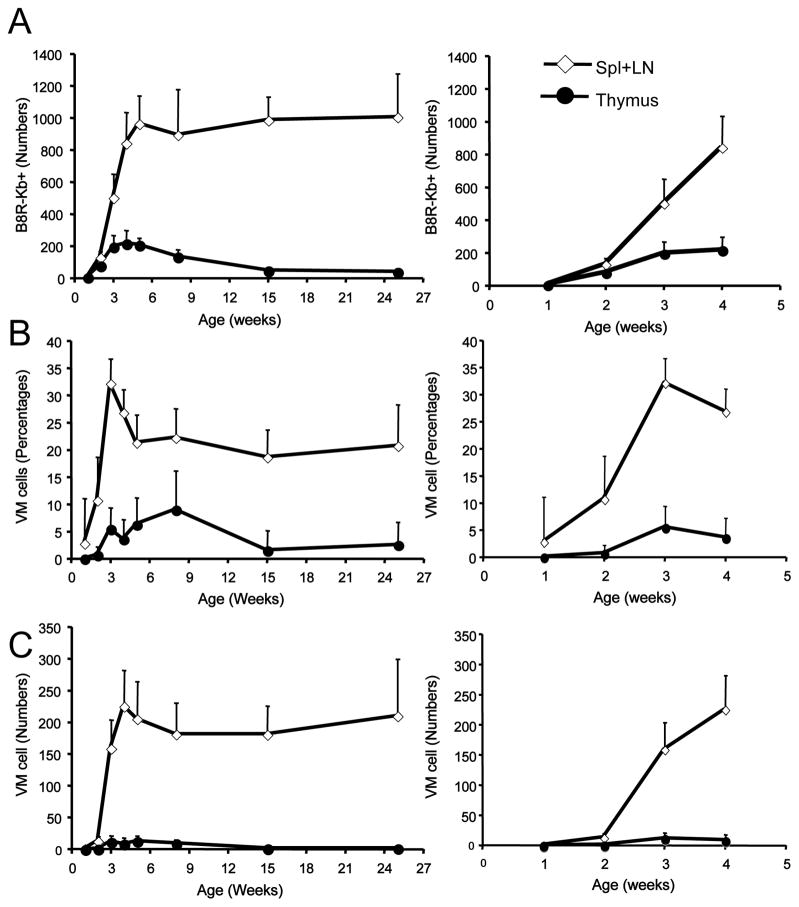
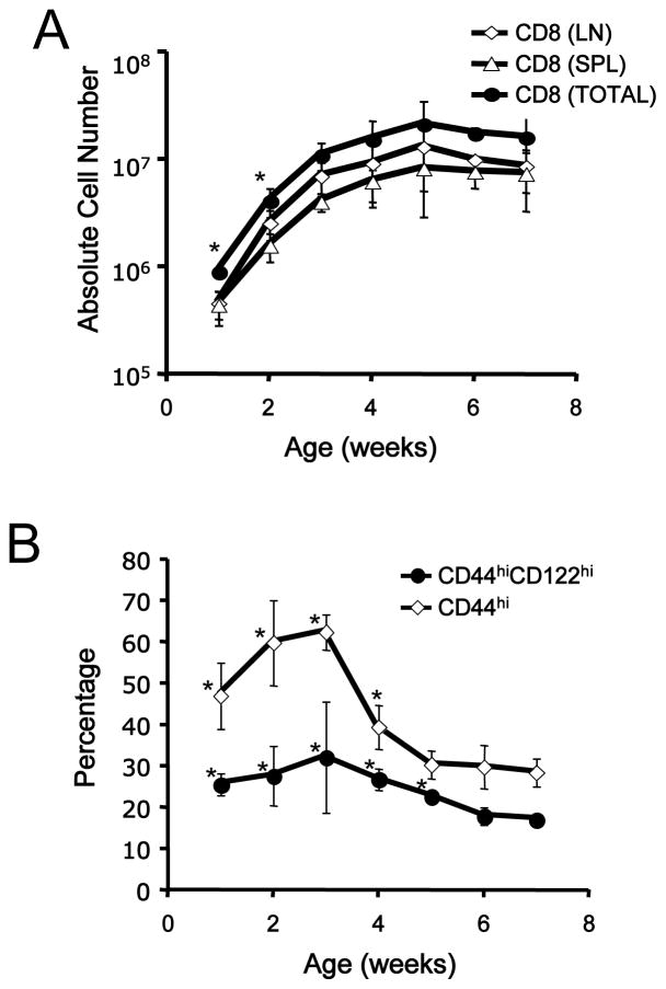
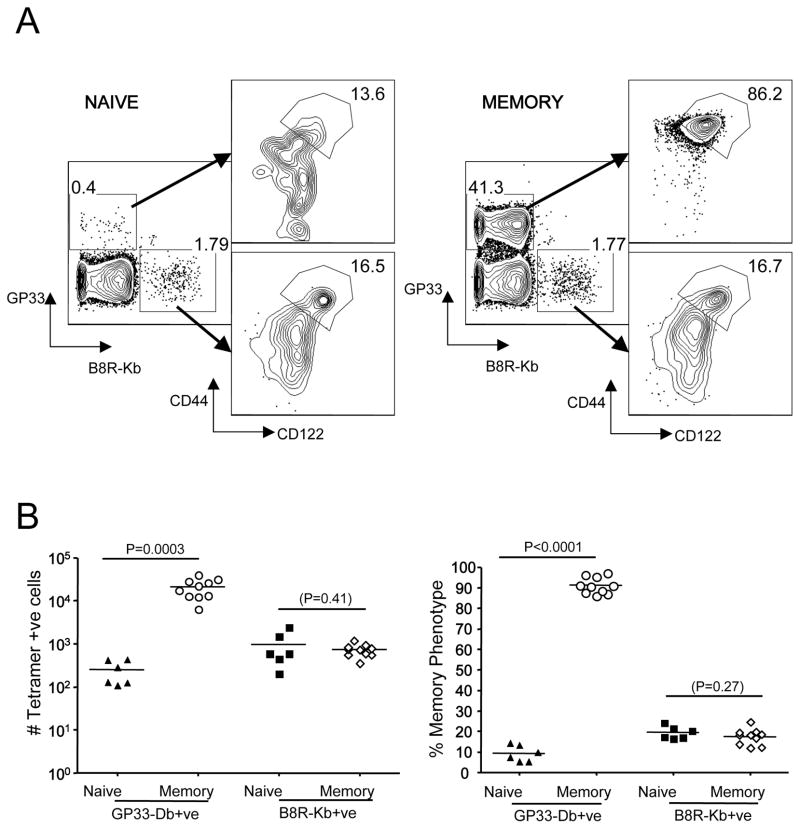
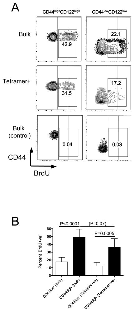
Memory CD8(+) T cells are an important component of the adaptive immune response against many infections, and understanding how Ag-specific memory CD8(+) T cells are generated and maintained is crucial for the development of vaccines. We recently reported the existence of memory-phenotype, Ag-specific CD8(+) T cells in unimmunized mice (virtual memory or VM cells). However, it was not clear when and where these cells are generated during normal development, nor the factors required for their production and maintenance. This issue is especially pertinent given recent data showing that memory-like CD8 T cells can be generated in the thymus, in a bystander response to IL-4. In this study, we show that the size of the VM population is reduced in IL-4R-deficient animals. However, the VM population appears first in the periphery and not the thymus of normal animals, suggesting this role of IL-4 is manifest following thymic egress. We also show that the VM pool is durable, showing basal proliferation and long-term maintenance in normal animals, and also being retained during responses to unrelated infection.


Figures

Figure 1
Figure 1. VM cells are detected among diverse foreign-antigen specific precursor pools
Peptide MHC-I tetramer-based enrichment was performed on spleen and lymph node cells from unprimed B6 mice using PE and APC-coupled tetramers representing five epitopes from MCMV (M38/Kb, M45/Db, M57/Kb, M139/Kb, and IE-3/Kb) and one from vaccinia virus (B8R/Kb). (A) shows the number of tetramer positive CD8+ T cells in the bound fraction. (B) represents the percentage of each tetramer positive CD8+ T pool that are either memory (CD44hi/CD122+) or naïve (CD44lo/dCD122−). The results in A and B and are compiled from at least 3 individual experiments.
Figure 2
Figure 2. VM cells are not dual TCR reactive T cells
Cells from lymph nodes and spleen were obtained from TCRα +/− B6 and wild type B6 mice, and stained for CD8, CD44, and CD122. Magnetic enrichment was performed using B8R/Kb tetramers. (A) Contour plots of both bulk CD8 T cells and B8R/Kb positive CD8+ T cells. Numbers represent the percentage of cells with the phenotype of memory (CD44hi/CD122+). (B) Depicts the number of B8R/Kb positive CD8+ T cells in the spleen and lymph nodes of TCRα +/− and B6 mice. (C) Shows the percentage of VM cells among the B8R/Kb positive CD8+ T cells of TCRα +/− and B6 mice. A total of 3 experiments were performed, each with 3 TCRα heterozygous B6 mice and 2 wild type B6 controls.
Figure 3
Figure 3. The size of the VM pool depends on IL-4 reactivity
Cells from lymph nodes and spleen were obtained from 9–10-week-old IL-4R−/− B6 and wild-type B6 mice, and stained for CD8, CD44, and CD122. MHC-I tetramer enrichment was also performed using B8R/Kb and HSVgB/Kb tetramers. (A) Depicts the number of B8R/Kb and HSVgB/Kb tetramer positive CD8+ T cells in the spleen and lymph nodes of IL-4R−/− and B6 mice. (B) Shows the percentage of memory phenotype cells among total, B8R/Kb and HSVgB/Kb tetramer positive CD8+ T cells in adult IL-4R−/− and B6 mice. A total of 3 experiments were performed, each with 3 IL-4R−/− B6 and 2 wild-type B6 control mice. In (C), 4–5-week-old wild type or IL-4R−/− mice were analyzed for frequency of memory-phenotype cells among CD8 single positive T cells from spleen and thymus.
Figure 4
Figure 4. VM cells arise first in the periphery during the neonatal period
Thymus and pooled spleen/lymph nodes were harvested from mice ranging from 1- to 25-weeks of age and tetramer enrichment was performed using B8R/Kb tetramers. CD8 single positive/CD3+ve cells were gated on. (A) Shows the number of B8R/Kb tetramer binding CD8+ T cells, (B) the percentage of memory-phenotype cells within this population and (C) shows the absolute number of memory-phenotype B8R/Kb tetramer binding CD8+ T cells in the thymus and pooled spleen and lymph nodes. For each group, the right-hand panel highlights the data from animals of 1- to 4-week old. Each time point represents a minimum of 2 experiments with at least 4 mice per experiment.
Figure 5
Figure 5. The frequency of memory-phenotype CD8 T cells is elevated during establishment of the peripheral T cell pool
Lymph node and spleen cells were obtained from B6 mice ranging from 1- to 7-week-old and stained with antibodies to CD4, CD8, CD44, and CD122. (A) shows the absolute number of bulk CD8+ T cells in the spleen, lymph nodes, and both tissues combined, while (B) shows the percentage of splenic CD8+ T cells that are of memory-phenotype (CD44hi/CD122+). These data derive from at least 2 individual experiments for each time point. An asterisk (*) indicates that a data set is significantly different (p<0.05) compared to the data from 7-week old animals.
Figure 6
Figure 6. VM cells are maintained during bystander immunization
Wild type B6 mice were infected with LCMV (Armstrong). Splenocytes were harvested 30–45 days later from these animals (“memory”) or uninfected controls (“naïve”) and subjected to enrichment using both B8R/Kb and GP33/Db tetramers. Numbers in the left-hand contour plots represent the percent of CD8 T cells in the bound fraction that are GP33/Db andB8R/Kb positive, while numbers in the right-hand contour plots represent the percent of VM cells for each specificity. These plots are representative of 3 experiments with a total of 10 LCMV memory mice and 6 unprimed B6 controls.
Figure 7
Figure 7. Basal proliferation of VM cells at steady state
Normal adult B6 mice were labeled with BrdU for 14–16 days, or were maintained in parallel without BrdU exposure (control). Spleen and lymph nodes cells were isolated and stained for cell surface markers and intracellular BrdU (for the “bulk” population), or were first subjected to tetramer enrichment (using a cocktail of B8R/Kb, M57/Kb and HSVgB/Kb tetramers) prior to surface and intracellular staining with antibodies. Events were gated on bulk or tetramer+ve naïve- and memory-phenotype CD8 T cells, as indicated. (A) shows representative data. Vertical blocks are overlaid on the contour plots to highlight the different levels of BrdU staining on naïve and memory T cells. The numbers on the plots indicate the percentage of total BrdU+ve cells (regardless of staining intensity). (B) shows compiled data on the percentage of BrdU+ve cells among the naïve and memory-phenotype cells among bulk and tetramer+ve CD8 T cells. Data are from 3 experiments deriving from two independent BrdU labeling cohorts.

Similar articles

Cited by

References

    1. Jameson SC, Masopust D. Diversity in T cell memory: an embarrassment of riches. Immunity. 2009;31:859–871. - PMC - PubMed
    1. Badovinac VP, Harty JT. CD8(+) T-cell homeostasis after infection: setting the ‘curve’. Microbes and infection/Institut Pasteur. 2002;4:441–447. - PubMed
    1. D’Cruz LM, Rubinstein MP, Goldrath AW. Surviving the crash: transitioning from effector to memory CD8+ T cell. Seminars in immunology. 2009;21:92–98. - PMC - PubMed
    1. Kaech SM, Ahmed R. Memory CD8+ T cell differentiation: initial antigen encounter triggers a developmental program in naive cells. Nature immunology. 2001;2:415–422. - PMC - PubMed
    1. Goldrath AW, Bevan MJ. Low-affinity ligands for the TCR drive proliferation of mature CD8+ T cells in lymphopenic hosts. Immunity. 1999;11:183–190. - PMC - PubMed

Publication types