. 2012 Feb 14;109(7):2302-7.
doi: 10.1073/pnas.1106799109. Epub 2012 Jan 30.

Src tyrosine kinase phosphorylation of nuclear receptor HNF4α correlates with isoform-specific loss of HNF4α in human colon cancer

Affiliations

Src tyrosine kinase phosphorylation of nuclear receptor HNF4α correlates with isoform-specific loss of HNF4α in human colon cancer

Karthikeyani Chellappa et al. Proc Natl Acad Sci U S A. .

Abstract

Src tyrosine kinase has long been implicated in colon cancer but much remains to be learned about its substrates. The nuclear receptor hepatocyte nuclear factor 4α (HNF4α) has just recently been implicated in colon cancer but its role is poorly defined. Here we show that c-Src phosphorylates human HNF4α on three tyrosines in an interdependent and isoform-specific fashion. The initial phosphorylation site is a Tyr residue (Y14) present in the N-terminal A/B domain of P1- but not P2-driven HNF4α. Phospho-Y14 interacts with the Src SH2 domain, leading to the phosphorylation of two additional tyrosines in the ligand binding domain (LBD) in P1-HNF4α. Phosphomimetic mutants in the LBD decrease P1-HNF4α protein stability, nuclear localization and transactivation function. Immunohistochemical analysis of approximately 450 human colon cancer specimens (Stage III) reveals that P1-HNF4α is either lost or localized in the cytoplasm in approximately 80% of tumors, and that staining for active Src correlates with those events in a subset of samples. Finally, three SNPs in the human HNF4α protein, two of which are in the HNF4α F domain that interacts with the Src SH3 domain, increase phosphorylation by Src and decrease HNF4α protein stability and function, suggesting that individuals with those variants may be more susceptible to Src-mediated effects. This newly identified interaction between Src kinase and HNF4α has important implications for colon and other cancers.


Conflict of interest statement

The authors declare no conflict of interest.

Figures

Fig. 1.
Fig. 1.
Src preferentially phosphorylates P1-HNF4α2 on Tyr14, Tyr 277, and Tyr279. (A) In vitro Src kinase assay with HNF4α IP’d from bacterial lysate containing full-length GST-tagged P1- or P2-HNF4α (α2 and α8, respectively), recombinant Src kinase and 32P-γ-ATP. Shown is an autoradiograph of a blot following SDS-PAGE. Bottom, IB with HNF4α Ab. (B) In vivo Src kinase assay of COS-7 cells cotransfected with HNF4α2 and c-Src WT or Y530F and treated 100 μM PV for different time points as indicated. IP’d HNF4α was probed with anti-pY (4G10) or HNF4α Ab. (C) Mass spectrometry (nano-LC/MS/MS) analysis of HNF4α IP’d from HEK293 cells cotransfected with c-Src Y530F and HNF4α2 or HNF4α8 after a 10-min PV treatment. A peptide containing Y277/Y279 was detected at m/z 1471.2, +2, corresponding to a doubly phosphorylated fragment. The identity of the 1471.2, +2 peptide was verified using a synthetic peptide corresponding to the peptide sequence shown. (D) In vivo Src kinase assay as in (B) but with WT or Y to F mutants of HNF4α2 cotransfected with c-Src Y530F into HEK293 cells and treated with 100 μM PV for 10 min prior to lysis. pY14, phospho-specific Ab to pY14 HNF4α.
Fig. 2.
Fig. 2.
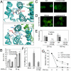
Tyr277 and Tyr279 are key residues for Src-mediated effects on HNF4α. (A) Three-dimensional model of human HNF4α LBD in the region of Y277 and Y279. The two subunits of the homodimer are shown in cyan and green. Key residues and helices are indicated. (B) Luciferase activity (relative light units, RLU) from transiently transfected Cos-7 cells with WT or double mutants of HNF4α2 and an HNF4α-responsive luciferase construct (ApoB.-85-47.E4.Luc). Data are the means of triplicate samples from one experiment; error bars show s.d. *P < 0.002 WT vs. Y277E/Y279E mutant. (C) Subcellular localization of WT and double mutants of Y277 and Y279 in HNF4α2 expressed in COS-7 cells. Immunolabeled cells (α445 Ab) were visualized using a Zeiss 510 confocal microscope (40×) and digitally magnified. (D) Subcellular localization as in (C) but after leptomycin B (LMB) or vehicle treatment for 8 h of HNF4α2 Y277E/ Y279E mutant. Immunolabeled cells (α445 Ab) were visualized with a Nikon Eclipse Ti inverted microscope (20×) and digitally magnified. (E) Fold difference (normalized RLU in the presence or absence of HNF4α2) of COS-7 cells transfected with WT or Y277E/Y279E mutant of HNF4α2 and ApoAI-4.Luc reporter treated with LMB or vehicle for 9 h. Data are the means of triplicate samples from one experiment; error bars show s.d. *P < 0.05 vehicle vs. LMB for WT, n.s., no significant change. (F) Fold difference (normalized RLU in the presence or absence of PGC1α) of WT or Y277E/Y279E mutant of HNF4α2 cotransfected in HEK293 cells with ApoAI-4.Luc reporter and CMV.β-gal. *P < 0.0005 fold difference in mock-transfected vs. WT HNF4α2. (G) Quantification of protein stability of WT and double mutants of Y277 and Y279 in HNF4α2 expressed in Cos-7 cells in the presence of 50 μM CHX at the indicated time points. Experiments in BG were performed two to three times with similar results.
Fig. 5.
Fig. 5.
Src SH3 domain interacts with proline-rich F domain of HNF4α; SNPs in HNF4α2 LBD and F domain affect Src-mediated phosphorylation. (A) Pulldown assays with GST.Src-SH3 protein and whole cell extract from HEK293 cells transfected with WT or the indicated SNP variants of HNF4α2. (B) In vivo Src kinase assay of HEK293 cells cotransfected with HNF4α2 and either c-Src Y530F or Src SH3 mutant (c-Src W121A/Y530F). IP’d HNF4α was probed with 4G10, pY14, or P1/P2 HNF4α Ab. (C) In vivo Src kinase assay as in Fig. 1B but with HNF4α2 WT or the indicated SNP variants cotransfected with c-Src Y530F into HEK293 cells. Right, quantification of the pY14 signal relative to the total HNF4α signal from one of three representative experiments. (D) Protein stability of WT and SNP variants of HNF4α2 cotransfected with c-Src Y530F into HEK293 cells. Shown is the average of triplicates from one of two independent experiments of the HNF4α IB signal normalized to Coomassie stain. *P < 0.02 SNP variants vs. WT. (E) Transcriptional activity of WT or SNP variants of HNF4α2 either in the presence or absence of c-Src Y530F in HEK293 cells cotransfected with pZL.HIV.LTR.AI-4 reporter and CMV.βgal. Shown is the RLU normalized to HNF4α protein and β-gal activity for WT (Left) and % of WT (set to 100%) for SNP variants (Right). Data are the mean + /-s.e.m. of two independent experiments performed in triplicates. *P < 0.05 for SNP variants in the presence vs. the absence of c-Src Y530F.
Fig. 3.
Fig. 3.
Active Src decreases P1- but not P2-HNF4α protein stability; tyrosine phosphorylation of endogenous P1-HNF4α. (A) Left, Protein stability of WT HNF4α2 expressed in HEK293 cells in the presence or absence of c-Src Y530F. Graph represents HNF4α protein level normalized to Coomassie stain. Data are means of triplicates from one of at least 10 representative experiments; error bars show s.e.m. *P < 0.05.Right top, Right, IB of HNF4α2 and β-actin from whole cell extracts of HEK293 treated with either DMSO or 50 μM MG132 for 8 h. Shown are representative blots from three independent experiments. (B) Protein stability of P1- and P2-HNF4α in CaCo2 cells harvested at the indicated time points after EGF + CHX treatment. Shown is one representative quantification of EGF-treated versus untreated controls from five independent experiments. (C) In vivo phosphorylation of endogenous HNF4α from CaCo2 cells. Top, IP’d phospho-Tyr proteins (4G10) were IB’d with either P1- or P2-HNF4α Abs. Bottom, IP’d HNF4α (α445) was probed with pY14 or P1-HNF4α Ab. (D) Mass spectrometry analysis as in Fig. 1C but of P1-HNF4α IP’d (αN1.14 Ab) from CaCo2 cells. Signals corresponding to the nonphosphorylated Y277/Y279 (m/z 1391.2, +2) and pY277pY279 (m/z 1471.2, +2) peptides were detected. Ratio of the amount of pY277/pY279 to Y277/Y279 is indicated. (E) As in (C, Top) but from CaCo2 cells treated with ethanol or 10 μM PP2 for different time points. Values corresponding to normalized phospho P1-HNF4α (IP’d P1-HNF4α divided by input P1-HNF4α) are shown. (F) IB of P1-HNF4α and P2-HNF4α from whole cell extracts of CaCo2 cells after 24 h or 48 h treatment with 10 μM PP2 or DMSO. Shown are triplicates from one of three independent experiments.
Fig. 4.
Fig. 4.
Src SH2 domain interacts with pY14 and is critical for phosphorylation of Y277 and Y279. (A) Pulldown assay with GST or GST.Src-SH2 proteins and whole cell extracts from HEK293 cells cotransfected with c-Src Y530F and WT or the indicated Y to F mutants of HNF4α2. IBs with α445 show the amount of HNF4α in the Input and after the Pulldown. (B) As in (A) but with human HNF4α2 and HNF4α8. (C) Pulldown assays as in (A) but with WT or R178A mutants of Src SH2 and 5 μg of pY14 peptide (Fig. S2D). Shown is an IB of bound peptide after Pulldown spotted onto PVDF membrane and detected by an Ab that recognizes the pY14 peptide. (D) Pulldown as in (A) but the interaction between WT HNF4α2 and GST.Src-SH2 was competed with either the pY14 or the Y14 peptide. Shown is the IB with the HNF4α antibody from one representative experiment and quantification of bound HNF4α relative to no peptide from 5 and 2 independent experiments for pY14 and Y14 competition, respectively. Error bar represents s.e.m. *P < 0.01 relative to no peptide competition. (E) In vivo Src kinase assay in HEK293 cells cotransfected in 100-mm plates with HNF4α2 WT (5 μg) and either c-Src Y530F or Src SH2 mutant (c-Src R178A/Y530F) at different ratios of transfected DNA (5 μg to 0.5 μg). IP’d HNF4α was probed with 4G10, pY14 or P1/P2 HNF4α Ab. Indicated is the fold difference in the phosphorylation signal with the Src SH2 mutant relative to c-Src Y530F at each ratio. Fold difference for the 1∶0.2 ratio was calculated using a darker exposure of the 4G10 blot. NA, not applicable as no signal was observed.
Fig. 6.
Fig. 6.
Preferential loss and cytoplasmic staining of P1-HNF4α in human colon cancer correlates with active Src; model of multistep phosphorylation of HNF4α by Src. (A) Diverse patterns of nuclear and cytoplasmic accumulation of HNF4α protein as detected by the P1-specific HNF4α Ab (brown staining) in colon cancers from different patients: a) total absence of P1-HNF4α in malignant epithelial cells; b) predominantly cytoplasmic with many nuclei negative for P1-HNF4α; c) cytoplasmic and nuclear P1-HNF4α; and d) strong nuclear with almost no cytoplasmic presence of P1-HNF4α. Magnification 200×. (B) Number of tumors with a given staining pattern of P1-HNF4α (a, b, c or d) shown in (A) out of the total cohort of 405. (C) Representative stains for P1-HNF4α and active Src (pSrc) in a subset of tumors: P1-HNF4α (I, III); pSrc (II, IV). (D, E) Model of multistep phosphorylation of P1-HNF4α by Src tyrosine kinase and effect of SNP’s in the HNF4α2 LBD (L280F) and F domain (P421L, P436S).

References

    1. Walther A, et al. Genetic prognostic and predictive markers in colorectal cancer. Nat Rev Cancer. 2009;9:489–499. - PubMed
    1. ACS. Colorectal Cancer Facts & Figures 2008-2010. Atlanta: American Cancer Society; 2008.
    1. Yeatman TJ. A renaissance for SRC. Nat Rev Cancer. 2004;4:470–480. - PubMed
    1. Iravani S, et al. Elevated c-Src protein expression is an early event in colonic neoplasia. Lab Invest. 1998;78:365–371. - PubMed
    1. Cartwright CA, Meisler AI, Eckhart W. Activation of the pp60c-src protein kinase is an early event in colonic carcinogenesis. Proc Natl Acad Sci USA. 1990;87:558–562. - PMC - PubMed

Publication types

LinkOut - more resources