. 2012 Feb 7;109(6):1979-84.
doi: 10.1073/pnas.1108705109. Epub 2012 Jan 20.

Active transcription and essential role of RNA polymerase II at the centromere during mitosis

Affiliations

Active transcription and essential role of RNA polymerase II at the centromere during mitosis

F Lyn Chan et al. Proc Natl Acad Sci U S A. .

Abstract

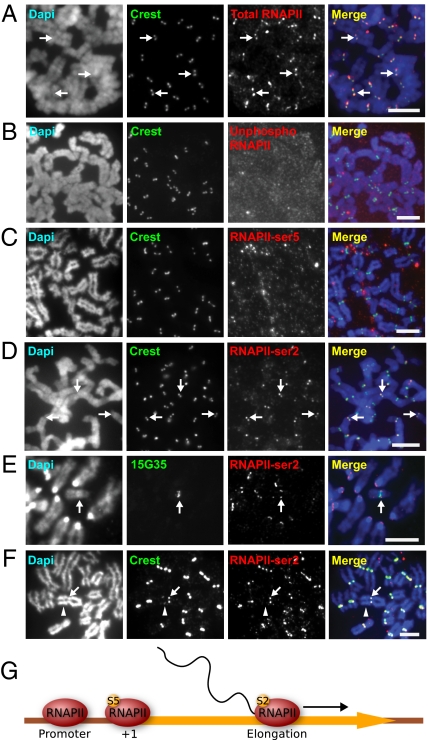
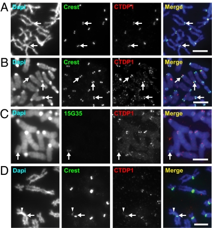
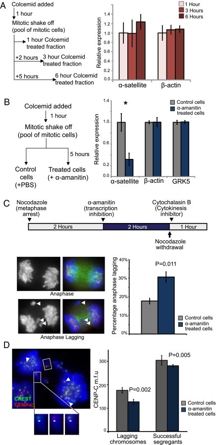
Transcription of the centromeric regions has been reported to occur in G1 and S phase in different species. Here, we investigate whether transcription also occurs and plays a functional role at the mammalian centromere during mitosis. We show the presence of actively transcribing RNA polymerase II (RNAPII) and its associated transcription factors, coupled with the production of centromere satellite transcripts at the mitotic kinetochore. Specific inhibition of RNAPII activity during mitosis leads to a decrease in centromeric α-satellite transcription and a concomitant increase in anaphase-lagging cells, with the lagging chromosomes showing reduced centromere protein C binding. These findings demonstrate an essential role of RNAPII in the transcription of α-satellite DNA, binding of centromere protein C, and the proper functioning of the mitotic kinetochore.


Conflict of interest statement

The authors declare no conflict of interest.

Figures

Fig. 1.
Fig. 1.
RNAPII-ser2 localizes to the mitotic kinetochore and is associated with active kinetochore activity. (A) Antibody 4H8total RNAPII immunostained the kinetochores of HeLa cells, as shown by the colocalization with CREST anti-centromere signals (arrows). (B) Antibody 8WG16unphosphorylated, specific for unphosphorylated RNAPII did not immunostain HeLa mitotic kinetochores. (C) Antibody H14phosphoSer5, which recognizes RNAPII phosphorylated at serine 5 (RNAPII-ser5) associated with transcription initiation, showed no staining of the kinetochores. (D) H5phosphoSer2, specific for elongating RNAPII, stained the kinetochores of mitotic HeLa cells (arrows). (E) Combined immunofluorescence/DNA-FISH (antibody H5phosphoSer2 and 10q25 band-specific 153G5 BAC probe) of mouse ES mardel (10) cells showed the presence of RNAPII-ser2 at the mardel (10) neocentromere (arrow) as well as at endogenous mouse kinetochores. (F) RNAPII-ser2 was present only at the active neocentromere of the PD-NC4 chromosomes (arrow) but not at the inactive native centromere (arrowhead, as indicated by the weaker CREST signals attributed to residual CENP-B). (G) Schematic depicting the change in RNAPII phosphorylation status across the transcription cycle. At the promoter, RNAPII is unphosphorylated. At transcription initiation, RNAPII is phosphorylated at serine 5 (S5), but before the RNAPII complex is competent for transcription elongation it must be phosphorylated at serine 2 (S2). (Scale bars: 5 μm.)
Fig. 2.
Fig. 2.
Transcription factor CTDP1 is found at the kinetochores of human and mouse cells as well as at neocentromeres. (A and B) CTDP1 localized to the kinetochores of mitotic human HeLa and mouse NIH 3T3 cells respectively, as evidenced by colocalization with CREST signals (arrows). (C) CTDP1 also localized to the kinetochore of the mardel (10) neocentromere, as shown by the colocalization of CTDP1 and 153G5 BAC DNA-FISH probe specific for the mardel (10) 10q25 neocentromere (arrow). (D) CTDP1 is enriched at the kinetochore of the active PD-NC4 neocentromere, as shown by its colocalization with the brighter CREST signal (arrow), but not at the inactive native alphoid centromere with reduced CREST signal (arrowhead). (Scale bars: 5 μm.)
Fig. 3.
Fig. 3.
The kinetochore is transcriptionally active during mitosis. (A) In situ transcription assay revealed transcriptional activity in cytospun NIH 3T3 interphase cells, as shown by nuclear and nucleolar FITC-rUTP incorporation (arrow). (B) The in situ transcription assay showed FITC-rUTP incorporation at the kinetochore regions (arrows) of the mitotic chromosomes. (C) FITC-rUTP incorporation at the kinetochore is sensitive to the RNAPII inhibitor α-amanitin. (D) CREST immunofluorescence performed after the in situ transcription assay showed that the FITC-rUTP signals colocalized with CREST signals (arrows). (E) Fluorescence intensity line scans of a single chromosome (boxed in D). Lateral (solid line) and axial (dashed line) fluorescence intensity line-scans through the kinetochores confirmed that the FITC-rUTP signals (green lines) colocalized with CREST signals (red lines); blue lines indicate DAPI staining. (Scale bars: 5 μm in AD, 2 μm in E.)
Fig. 4.
Fig. 4.
Transcription of α-satellite DNA during mitosis is mediated by RNAPII and is required for correct kinetochore function. (A) A mitotic shake-off protocol and real-time qRT-PCR were used to measure the levels of α-satellite RNA in metaphase-arrested cells. Briefly, 14ZBHT cells were treated with colcemid for 1 h, and mitotic shake-off was performed to isolate a population of mitotic-enriched cells (average mitotic index = 70%; n = 5). Cells were maintained in mitotic arrest for an additional 0, 2, or 5 h to give 1-, 3-, and 6-h mitotic-arrested populations. Total RNA was reverse-transcribed into cDNAs for real-time qRT-PCR using the ΔΔCT method (normalized against the mean of three housekeeping genes, β-actin, GAPDH, and HPRT). No significant change in the levels of α-satellite or actin transcripts was detected. (B) The effect of RNAPII inhibition on centromere transcription was assayed. A pool of mitotic 14ZBHT cells was isolated via mitotic shake-off and split into two fractions, an α-amanitin–treated fraction (incubated with 50 μg/mL α-amanitin) and a control fraction (PBS added), for a further 5 h. RT-PCR quantification was performed as described previously. α-Amanitin–mediated RNAPII inhibition caused a significant decrease in α-satellite levels (by 68%; n = 4; *P = 0.016; Student's t test). Transcripts of the control genes, G protein-coupled receptor kinase 5 and β-actin, were unaffected. (C) A protocol was designed to target RNAPII inhibition to mitotic cells, and an anaphase-lagging assay was used to detect kinetochore dysfunction. 14ZBHT cells were treated with nocodazole for 2 h to enrich for mitotic cells; then cells were treated with α-amanitin (50 μg/mL) or PBS for a further 2 h. Cells then were released from mitotic arrest and placed in cytochalasin B-containing medium to inhibit cytokinesis. One hundred anaphase events were scored as normal or anaphase lagging. Arrowheads indicate examples of lagging chromosomes. α-Amanitin treatment caused a significant increase in lagging anaphases (n = 3; P = 0.011; Student's t test). (D) An anaphase-lagging cell. Lagging chromosomes are indicated by a box, and successfully segregated sister kinetochores are indicated by arrowheads. Insets show close-up images (CREST in green, CENP-C in red) (Left) and merged images (Right) of a lagging chromosome. The fluorescence intensities of CENP-C at the kinetochores of lagging chromosomes (an example is indicated by asterisk) and successfully segregated chromosomes were measured. After α-amanitin inhibition, the mean fluorescence intensity of CENP-C at the kinetochores of the successfully segregated chromosomes was reduced slightly (by 7%; P = 0.005); an even greater reduction of CENP-C was seen at the kinetochores’ lagging chromosomes (28%; P = 0.002; based on 77 control and 107 α-amanitin–treated chromosomes from three biological replicates). Error bars in AD represent SEM.

References

    1. Marshall OJ, Chueh AC, Wong LH, Choo KH. Neocentromeres: New insights into centromere structure, disease development, and karyotype evolution. Am J Hum Genet. 2008;82:261–282. - PMC - PubMed
    1. Ekwall K. Epigenetic control of centromere behavior. Annu Rev Genet. 2007;41:63–81. - PubMed
    1. Volpe TA, et al. Regulation of heterochromatic silencing and histone H3 lysine-9 methylation by RNAi. Science. 2002;297:1833–1837. - PubMed
    1. Kanellopoulou C, et al. Dicer-deficient mouse embryonic stem cells are defective in differentiation and centromeric silencing. Genes Dev. 2005;19:489–501. - PMC - PubMed
    1. May BP, Lippman ZB, Fang Y, Spector DL, Martienssen RA. Differential regulation of strand-specific transcripts from Arabidopsis centromeric satellite repeats. PLoS Genet. 2005;1:e79. - PMC - PubMed

Publication types

MeSH terms

LinkOut - more resources