. 2012 Feb 14;109(7):2336-41.
doi: 10.1073/pnas.1109526109. Epub 2012 Jan 27.

Aggregation of α-synuclein is kinetically controlled by intramolecular diffusion

Affiliations

Aggregation of α-synuclein is kinetically controlled by intramolecular diffusion

Basir Ahmad et al. Proc Natl Acad Sci U S A. .

Abstract

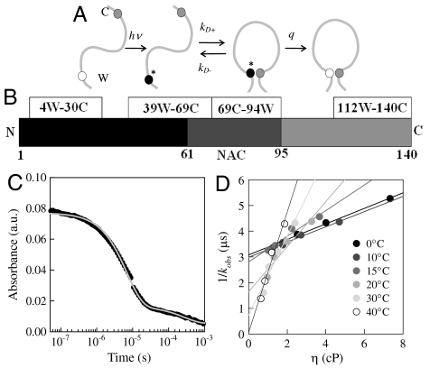
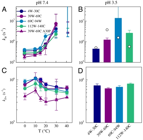
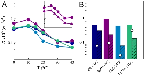
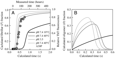
We hypothesize that the first step of aggregation of disordered proteins, such as α-synuclein, is controlled by the rate of backbone reconfiguration. When reconfiguration is fast, bimolecular association is not stable, but as reconfiguration slows, association is more stable and subsequent aggregation is faster. To investigate this hypothesis, we have measured the rate of intramolecular diffusion in α-synuclein, a protein involved in Parkinson's disease, under solvent conditions that accelerate or decelerate aggregation. Using the method of tryptophan-cysteine (Trp-Cys) quenching, the rate of intramolecular contact is measured in four different loops along the chain length. This intrinsically disordered protein is highly diffusive at low temperature at neutral pH, when aggregation is slow, and compacts and diffuses more slowly at high temperature or low pH, when aggregation is rapid. Diffusion also slows with the disease mutation A30P. This work provides unique insights into the earliest steps of α-synuclein aggregation pathway and should provide the basis for the development of drugs that can prevent aggregation at the initial stage.


Conflict of interest statement

The authors declare no conflict of interest.

Figures

Fig. 1.
Fig. 1.
(A) Determination of the rate of contact formation between the probe, tryptophan (W), and the quencher, cysteine (C), within an unfolded protein. Pulsed optical excitation leads to population of the lowest excited triplet state of tryptophan. Tryptophan contacts cysteine in a diffusion-limited process with rate kD+, and then either diffuses away or is quenched by the cysteine. (B) Sequence diagram of α-synuclein with positions of measured loops indicated. (C) Transient absorption of the tryptophan triplet state in the 112W-140C loop at pH 7.4, T = 0° C. (D) Observed lifetimes of the triplet state of 39W-69C loop at various temperatures and viscosities. The lines are independent fits of the data at each temperature.
Fig. 2.
Fig. 2.
Reaction-limited (A) and diffusion-limited (C) rates for various loops. (A and C) The rates versus temperature at pH 7.4. At T = 40° C, the dashes represent the lower limit of kR as determined by inverse error of the intercept. The diffusion-limited rates are calculated for the viscosity of water at each temperature. (B and D) The rates for all temperatures at pH 3.5. Because 1/kobs at pH 3.5 is temperature independent, plots of kR and kD+ versus T would be flat lines. The diffusion-limited rates are calculated for η = 1 cP (20° C in water). The points in B are reaction-limited rates calculated from the probability distributions shown in Fig. S9.
Fig. 3.
Fig. 3.
Diffusion coefficients calculated from measured kD+ and probability distributions of Trp-Cys distances calculated from the energy reweighted WLC model for (A) pH 7.4 at various temperatures and (B) pH 3.5 at all temperatures (hatched bars). D at pH 7.4 and T = 10° C are shown for comparison in B. The colors correspond to the different loops as designated in Fig. 2. The points are D calculated from probability distributions shown in Fig. S9 and the measured kD+ at pH 3.5 (Fig. 2D). (Inset) Comparison of diffusion coefficients between 39W-69C (circles) and 39W-69C A30P (triangles).
Fig. 4.
Fig. 4.
(A) Fibrillar fraction calculated from the model given in the text (state F) for 39W-69C at pH 7.4, T = 10° C (black line), pH 7.4, T = 40° C (dark gray line), 69C-94W at pH 3.5 (medium gray line), and 39W-69C A30P at pH 7.4, T = 40° C (light gray line) using the rates given in Table 1. The circles are experimental measurements of aggregation of mutant 69C-94W detected by ThT fluorescence (right and top axes) scaled to match the calculated rates (left and bottom axes). (B) Oligomer fraction (state O) calculated from the model using the same rates as A.

Similar articles

Cited by

References

    1. Spillantini MG, et al. α-Synuclein in Lewy bodies. Nature. 1997;388:839–840. - PubMed
    1. Goedert M. Alpha-synuclein and neurodegenerative diseases. Nat Rev Neurosci. 2001;2:492–501. - PubMed
    1. Wang W, et al. A soluble α-synuclein construct forms a dynamic tetramer. Proc Natl Acad Sci USA. 2011;108:17797–17802. - PMC - PubMed
    1. Bartels T, Choi JG, Selkoe DJ. α-Synuclein occurs physiologically as a helically folded tetramer that resists aggregation. Nature. 2011;477:107–110. - PMC - PubMed
    1. Bertoncini CW, et al. Release of long-range tertiary interactions potentiates aggregation of natively unstructured α-synuclein. Proc Natl Acad Sci USA. 2005;102:1430–1435. - PMC - PubMed

Publication types

Substances