. 2012 Feb 7;109(6):2126-31.
doi: 10.1073/pnas.1111715109. Epub 2012 Jan 23.

Modulation of value representation by social context in the primate orbitofrontal cortex

Affiliations

Modulation of value representation by social context in the primate orbitofrontal cortex

João C B Azzi et al. Proc Natl Acad Sci U S A. .

Erratum in

  • Proc Natl Acad Sci U S A. 2012 Mar 6;109(10):4020

Abstract

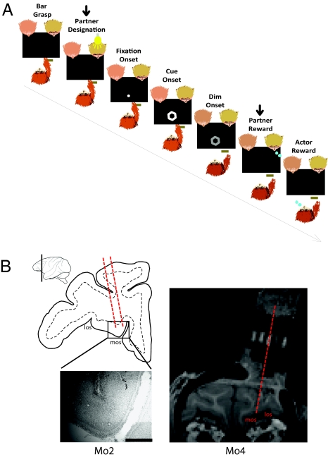
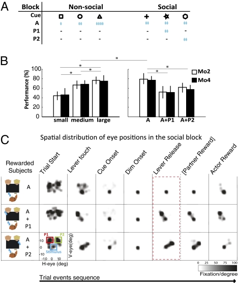
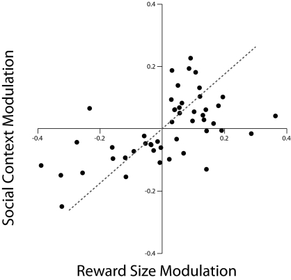
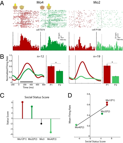
Primates depend for their survival on their ability to understand their social environment, and their behavior is often shaped by social circumstances. We report that the orbitofrontal cortex, a brain region involved in motivation and reward, is tuned to social information. Macaque monkeys worked to collect rewards for themselves and two monkey partners. Behaviorally, monkeys discriminated between cues signaling large and small [corrected] rewards, and between cues signaling rewards to self only and reward to both self and another monkey, with a preference for the former over the latter in both instances. Single neurons recorded during this task encoded the meaning of visual cues that predicted the magnitude of future rewards, as well as the motivational value of rewards obtained in a social context. Furthermore, neuronal activity was found to track momentary social preferences and partner's identity and social rank. The orbitofrontal cortex thus contains key neuronal mechanisms for the evaluation of social information.


Conflict of interest statement

The authors declare no conflict of interest.

Figures

Fig. 1.
Fig. 1.
Task description and localization of recording sites in the OFC. (A) Typical joint-reward trial sequence in which a monkey responds to a visual instruction by releasing a manual lever to obtain a reward for itself and for a predesignated partner. The shape of the reward cue specifies the trial's reward contingency. The arrow above the panels indicates the steps that are omitted on self-only reward trials. (B, Left) Drawing of a coronal section through the left frontal lobe of monkey Mo2 at the level indicated on the lateral view. Marks left by two electrode tracks are visible on the enlarged Nissl-stained picture. (Scale bar, 1 mm.) At the level shown in this drawing, area 13 is roughly bounded laterally by the fundus of the lateral orbital sulcus (LOS) and medially by the fundus of the medial orbital sulcus (MOS). (Right) Coronal view of anatomical MRI scans of monkey Mo4 showing the recording chamber position. A special grid with five holes in the central row filled with omega 3 and spaced 3-mm apart was inserted in the chamber, serving as reference to identify the position and angle of electrode tracks with respect to the underlying cortical tissue.
Fig. 2.
Fig. 2.
Behavioral performance. (A) Reward configurations on nonsocial and social trial blocks. (B) Mean percent correct responses of monkeys Mo2 (n = 47 sessions) and Mo4 (n = 45 sessions) as a function of reward size in the nonsocial block, and for joint and self-only reward conditions in the social block (asterisks and horizontal lines indicate significant pair-wise comparisons, P < 0.01). (C) Distribution of eye fixations over the workspace for different task epochs (combined data from Mo2 and Mo4). On joint-reward trials, monkeys briefly looked at the designated partner's face before fixing their eyes on the visual cue. Right after responding to dimming of the cue, they shifted their gaze back to the partner's face, thereby anticipating the delivery of the partner's reward. On self-only reward trials, the monkeys kept looking at the screen until their own reward was delivered.
Fig. 3.
Fig. 3.
Single-unit and population activity of OFC neurons in the nonsocial and social blocks. (Left three panels in A and C) Examples of single units with, respectively, up-modulated and down-modulated responses as a function of reward size; (Right three panels) Activity of the same two neurons in the social block. (B and D) Normalized spike density curves and mean discharge rate for the neuron population from which the corresponding single-unit examples are drawn. The thick horizontal bar below the spike density curves in B indicates the time window used for computing all statistical tests on mean population activity (Right). The asterisks and thin black or blue horizontal lines indicate significant pair-wise comparisons (P < 0.01).
Fig. 4.
Fig. 4.
Relationship between reward size and social context modulation. Modulation indices of general form (ab)/(a + b) were computed for all cells exhibiting cue-related response in the nonsocial and social block (SI Experimental Procedures, Data Analysis). For indicative purposes, indices of 0.11, 0.20, and 0.33 correspond to activity differentials of 25%, 50%, and 100%, respectively (regression r2 = 0.329, P < 0.02).
Fig. 5.
Fig. 5.
Face-selective responses of OFC neurons. (A) Single unit from monkeys Mo4 (Left) and Mo2 (Right). Both cells respond more strongly to P1 than P2 (Left: P1= 62.4 sp/s and P2= 52.5 sp/s; Right: P1= 24.8 sp/s and P2= 9.5 sp/s). (B) Respective normalized population spike density curves and mean discharge rate. (C) Social status score of the four members of the colony (SI Experimental Procedures, Animals). (D) Relationship between face-selective mean population activity and social status score (regression r2 = 0.937, P < 0.002). Color codes correspond to the same individuals in the different panels. (Conventions as in Fig. 3, asterisk and horizontal lines indicate significant pair-wise comparisons, P < 0.05 or better).

Similar articles

Cited by

References

    1. Rolls ET, Hornak J, Wade D, McGrath J. Emotion-related learning in patients with social and emotional changes associated with frontal lobe damage. J Neurol Neurosurg Psychiatry. 1994;57:1518–1524. - PMC - PubMed
    1. Plassmann H, O'Doherty J, Rangel A. Orbitofrontal cortex encodes willingness to pay in everyday economic transactions. J Neurosci. 2007;27:9984–9988. - PMC - PubMed
    1. Rolls ET. The orbitofrontal cortex and reward. Cereb Cortex. 2000;10:284–294. - PubMed
    1. Morrison SE, Salzman CD. The convergence of information about rewarding and aversive stimuli in single neurons. J Neurosci. 2009;29:11471–11483. - PMC - PubMed
    1. Tremblay L, Schultz W. Relative reward preference in primate orbitofrontal cortex. Nature. 1999;398:704–708. - PubMed

Publication types