. 2012 Feb 21;109(8):3083-8.
doi: 10.1073/pnas.1112729109. Epub 2012 Feb 1.

Pulmonary infection by Yersinia pestis rapidly establishes a permissive environment for microbial proliferation

Affiliations

Pulmonary infection by Yersinia pestis rapidly establishes a permissive environment for microbial proliferation

Paul A Price et al. Proc Natl Acad Sci U S A. .

Abstract

Disease progression of primary pneumonic plague is biphasic, consisting of a preinflammatory and a proinflammatory phase. During the long preinflammatory phase, bacteria replicate to high levels, seemingly uninhibited by normal pulmonary defenses. In a coinfection model of pneumonic plague, it appears that Yersinia pestis quickly creates a localized, dominant anti-inflammatory state that allows for the survival and rapid growth of both itself and normally avirulent organisms. Yersinia pseudotuberculosis, the relatively recent progenitor of Y. pestis, shows no similar trans-complementation effect, which is unprecedented among other respiratory pathogens. We demonstrate that the effectors secreted by the Ysc type III secretion system are necessary but not sufficient to mediate this apparent immunosuppression. Even an unbiased negative selection screen using a vast pool of Y. pestis mutants revealed no selection against any known virulence genes, demonstrating the transformation of the lung from a highly restrictive to a generally permissive environment during the preinflammatory phase of pneumonic plague.


Conflict of interest statement

The authors declare no conflict of interest.

Figures
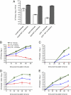
Fig. 1.
Fig. 1.
Wild-type Y. pestis is able trans-complement avirulent isogenic strains of Y. pestis. (A) Individual intranasal infections (white bars) for ΔyopH (YP373), Δpla (YP102), or pCD1 (YP6) mutants or matched coinfections (gray bars) with wild-type Y. pestis in C57BL/6J mice. CFU per lung were determined at 48 hpi using BHI agar plates containing X-Gal. All CFU differences between individual infections and corresponding coinfections (A–E, all time points) are statistically significant (P ≤ 0.01; unpaired t test). (B–E) Kinetics of infection for pCD1 mutants (YP6, red), wild-type (YP160, black), and a 1:1 coinfection of pCD1 mutants (blue) and wild-type Y. pestis (green) infected intranasally (B and C) or subcutaneously (D and E). CFU per tissue were determined at various time points of infection for lungs (B), spleen (C and E), and cervical lymph nodes (D). Each bar (A) or point (B–E) represents the mean CFU recovered from five mice. The limit of detection is represented by a dotted line. Data and statistical conclusions are representative of at least two independent experiments. Error bars represent SEM for the bar graph and SD from the mean for line graphs.
Fig. 2.
Fig. 2.
Relative numbers and distribution of virulent and avirulent Y. pestis during pulmonary coinfections. (A) Kinetics of infection for C57BL/6J mice intranasally infected with varying ratios of pCD1 mutants (YP6) and wild-type Y. pestis (YP160) [10:1 (black), 1:1 (blue), and 1:10 (orange) pCD1: wild-type]. CFU per lung were determined at various time points of infection. Each point represents the mean CFU recovered from five mice. Each ratio maintained a 1:10 mutant: wild-type ratio after 24 hpi. There were no statistically significant differences between the CFU of pCD1 mutants between any given ratio at each time point (P > 0.05; unpaired t test). The limit of detection is represented by a dotted line. Data and statistical conclusions are representative of at least two independent experiments. Error bars represent SD from the mean. (B) Mice were coinoculated intranasally with gfp-expressing pCD1 mutants (BGY19) and rfp-expressing wild-type Y. pestis (YP337). At 48 hpi, lungs were fixed and cryosectioned. Fluorescence deconvolution images were generated from 10-μm sections. The image is representative of multiple independent experiments. (Scale bar, 10 μm.)
Fig. 3.
Fig. 3.
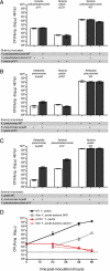
Specificity of bacterial trans-complementation during pulmonary infection. (A) Y. pseudotuberculosis, which harbors the same T3SS as Y. pestis, is unable to trans-complement in the lung. Individual intranasal infections (white bars) for pYV Y. pseudotuberculosis IP2666 mutants (BGY39), pCD1 Y. pestis mutants (BGY14-1), and wild-type Y. pseudotuberculosis IP2666 (BGY30) and matched coinfections (gray bars) with wild-type Y. pseudotuberculosis IP2666 in C57BL/6J mice (all differences between individual and co-infections were not significant, P > 0.05; unpaired t test). (B) K. pneumoniae, another pulmonary pathogen, is unable to trans-complement in the lung. Individual intranasal infections (white bars) for ΔcpsB K. pneumoniae mutants (VK060), pCD1 Y. pestis mutants (BGY14-1) and wild-type K. pneumoniae (KPPR1), and matched coinfections (gray bars) with wild-type K. pneumoniae in C57BL/6J mice (all differences between individual and co-infections were not significant, P > 0.05; unpaired t test). (C) Y. pestis is able to trans-complement avirulent strains of other pathogens. Individual intranasal infections (white bars) for ΔcpsB K. pneumoniae mutants (VK060), pYV Y. pseudotuberculosis IP2666 mutants (BGY38) and wild-type Y. pestis (YP160), and matched coinfections (gray bars) with wild-type Y. pestis in C57BL/6J mice (all differences between individual and co-infections were significant, P ≤ 0.001; unpaired t test). (D) Wild-type Y. pestis creates a permissive environment that allows nonpathogenic bacteria to proliferate in the lung. Kinetics of infection for pCD1 mutants (red) and wild-type Y. pestis (black) in C57BL/6J mice infected intranasally. CFU per lung were determined at various time points of infection on both BHI (Y. pestis) and BHI-MOX agar plates (non-Y. pestis bacteria). Each bar (A–C) or point (D) represents the mean CFU recovered from five mice. The limit of detection is represented by a dotted line. Data and statistical conclusions are representative of at least two independent experiments. Error bars represent SEM for bar graphs and SD from the mean for line graphs.

Similar articles

Cited by

References

    1. Bubeck SS, Cantwell AM, Dube PH. Delayed inflammatory response to primary pneumonic plague occurs in both outbred and inbred mice. Infect Immun. 2007;75:697–705. - PMC - PubMed
    1. Agar SL, et al. Characterization of a mouse model of plague after aerosolization of Yersinia pestis CO92. Microbiology. 2008;154(Pt 7):1939–1948. - PubMed
    1. Lathem WW, Crosby SD, Miller VL, Goldman WE. Progression of primary pneumonic plague: A mouse model of infection, pathology, and bacterial transcriptional activity. Proc Natl Acad Sci USA. 2005;102:17786–17791. - PMC - PubMed
    1. Koster F, et al. Milestones in progression of primary pneumonic plague in cynomolgus macaques. Infect Immun. 2010;78:2946–2955. - PMC - PubMed
    1. Montminy SW, et al. Virulence factors of Yersinia pestis are overcome by a strong lipopolysaccharide response. Nat Immunol. 2006;7:1066–1073. - PubMed

Publication types