. 2012 Feb 7;109(6):2096-101.
doi: 10.1073/pnas.1113775109. Epub 2012 Jan 20.

Host-targeting protein 1 (SpHtp1) from the oomycete Saprolegnia parasitica translocates specifically into fish cells in a tyrosine-O-sulphate-dependent manner

Affiliations

Host-targeting protein 1 (SpHtp1) from the oomycete Saprolegnia parasitica translocates specifically into fish cells in a tyrosine-O-sulphate-dependent manner

Stephan Wawra et al. Proc Natl Acad Sci U S A. .

Abstract

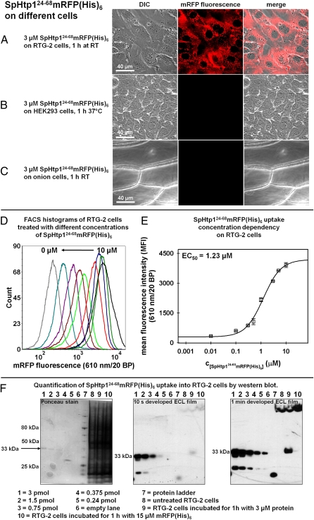
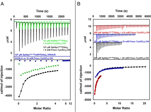
The eukaryotic oomycetes, or water molds, contain several species that are devastating pathogens of plants and animals. During infection, oomycetes translocate effector proteins into host cells, where they interfere with host-defense responses. For several oomycete effectors (i.e., the RxLR-effectors) it has been shown that their N-terminal polypeptides are important for the delivery into the host. Here we demonstrate that the putative RxLR-like effector, host-targeting protein 1 (SpHtp1), from the fish pathogen Saprolegnia parasitica translocates specifically inside host cells. We further demonstrate that cell-surface binding and uptake of this effector protein is mediated by an interaction with tyrosine-O-sulfate-modified cell-surface molecules and not via phospholipids, as has been reported for RxLR-effectors from plant pathogenic oomycetes. These results reveal an effector translocation route based on tyrosine-O-sulfate binding, which could be highly relevant for a wide range of host-microbe interactions.


Conflict of interest statement

The authors declare no conflict of interest.

Figures

Fig. 1.
Fig. 1.
The N-terminal leader peptide of SpHtp1 (amino acids 24–68) shows host cell-specific translocation. (A–C) The SpHtp1 leader fusion protein shows an autonomous translocation activity on the rainbow trout gonad cell line RTG-2 (A) but not with the human derived HEK293 cell line (B) or onion epidermis cells (C). Cells were incubated for 1 h with 3 μM SpHtp124-68mRFP(His)6 in L15-medium containing 10% FCS for the RTG-2 cells, in DMEM-medium (+10% FCS) for the HEK293 cells and in PBS containing 10% BSA for the onion epidermis cells at the indicated temperatures. Red channel: mRFP fluorescence; white channel: differential interference contrast (viability controls can be found in SI Appendix, Figs. S2 and S4). (D) Flow cytometry histograms for one repeat of the concentration dependency measurement. The RTG-2 cells were treated for 1 h at room temperature with either 10 μM (black), 5 μM (dark green), 3 μM (dark blue), 1 μM (red), 0.5 μM (green), 0.3 μM (brown), 0.1 μM (dark red), 0.01 μM (magenta), and 0 μM (gray) SpHtp124-68mRFP(His)6. From 10,000 counted events, a homogenous population of cells that were TO-PRO-3 iodide-negative was selected (on average ∼80% of all events) and further analyzed. (E) EC50 determination of SpHtp124-68mRFP(His)6 uptake into RTG-2 cells using the MFI from the FACS histograms (exemplarily shown in D). The errors are the SDs of the MFI values from three independent experiments. The obtained EC50 was 1.23 μM SpHtp124-68mRFP(His)6. (F) Quantification of SpHtp124-68mRFP(His)6 translocation into RTG-2 cells by Western blotting. Samples were prepared as described for FACS analyses then, following trypsination, the cells were harvested by centrifugation and resuspended 2× in PBS to remove excess trypsin. The wet cell pellets containing ∼2 × 10⁶ cells (∼20 μL) were lysed at 95 °C in 100 μL of Laemmli sample buffer containing 8 M urea, 2% β-mercaptoethanol, and 1 mM PMSF. Sample volume loaded was 15 μL. (Lanes 1–5) A total amount of 3 pmol (lane 1), 1.5 pmol (lane 2), 0.75 pmol (lane 3), 0.375 pmol (lane 4), and 0.24 pmol (lane 5) of purified SpHtp124-68mRFP(His)6 was loaded. Lane 6 was kept empty. Lane 8 contained a sample of RTG-2 cells incubated without protein, and lane 9 contained a RTG-2 cells incubated for 1 h with 3 μM SpHtp124-68mRFP(His)6. Lane 10 shows a sample incubated for 1 h with 15 μM mRFP(His)6. (Left) Ponceau stain of the proteins after transfer onto a nitrocellulose membrane. (Center and Right) ECL films after 10-s and 1-min development, respectively. The amount of SpHtp124-68mRFP(His)6 was determined by densitometric analysis of the band at 33 kDa and resulted in a value of ∼0.4 pmol.
Fig. 2.
Fig. 2.
The translocation activity of the SpHtp1 is dependent on sulfate-modified cell-surface molecules. (A–J) Confocal microscopy images of RTG-2 cells: Cells incubated for 1 h with 15 μM mRFP(His)6 (A) did not show any mRFP fluorescence inside the cells in contrast to cells incubated with 3 μM SpHtp124-68mRFP(His)6 (B). The mutated full-length SpHtp1-mRFP fusion protein, in which the KRHLR amino acids had been substituted with GGHLG, did not show any translocation into the cells under identical conditions (C). The reported RxLR-protein translocation inhibitor inositol-1,4-diphosphate (19) did not affect the translocation of SpHtp124-68mRFP(His)6 (D). However, pretreatment of the cells for 48 h with 70 mM of the sulfotransferase inhibitor NaClO3 (30) strongly inhibited the translocation of this protein (E). A similar effect was observed when the cells were 3-h preincubated with 1.1 U of a type VI aryl-sulphatase from Aerobacter aerogenes (34) (F). Incubation of the cells for 1 h with 10 μg/mL of an antityrosine-phosphate specific antibody before the application of SpHtp124-68mRFP(His)6 only slightly reduced the uptake of this protein (G). A much stronger effect was observed when an anti-tyrosine-sulfate specific antibody was used under identical conditions (H). Using the modified tyrosine amino acid H-Tyr(PO3)-OH at a concentration of 5 mM to compete for the SpHtp124-68mRFP(His)6 binding, the fluorescence levels found inside the cells were not significantly affected compared with the control (I). In contrast, with the same amount of tyrosine-sulfate [H-Tyr(SO3)-OH] a much stronger inhibitory effect was observed (J). Red channel: mRFP fluorescence; white channel: differential interference contrast (viability controls can be found in SI Appendix, Fig. S4). (K) Flow cytometric quantification of the mRFP-fluorescence uptake into RTG-2 cells corresponding to the images shown in A–J. Cells were grown in 25-cm2 flasks and washed 5× with L15 medium before incubation with the respective protein concentration dilute in L15-medium containing 10% FCS. After 1-h incubation, the cells were washed 3× 5 min with PB), 1× 10 min with PBS containing TO-PRO-3 iodide in a 1:1,000 dilution, 2× 5 min with PBS adjusted to pH 5.5, and 2× 5 min with PBS adjusted to pH 8.5. Subsequently, the cells were detached with 1 mL of 0.5 mg/mL trypsin (+1 mM EDTA), resulting in samples with ∼2 × 10⁶ cells/mL. From 10,000 events counted, a homogenous population of cell that were TO-PRO-3 iodide-negative was selected (on average ∼80% of all events) and further analyzed. Plotted are the MFI values averaged from three individual experiments. Error bars are the SD of the MFI values. Cells were incubated with 15 μM mRFP(His)6 (column 1), 3 μM SpHtp124-68mRFP(His)6 (column 2), 3 μM of the SpHtp124-198mRFP(His)6GGHLG mutant (column 3), or 3 μM SpHtp124-68mRFP(His)6 in combination with the indicated treatments (columns 5–10) or nontreated RTG-2 cells (column 11). Example raw data for one repeat can be found in the SI Appendix.
Fig. 3.
Fig. 3.
The N-terminal leader of SpHtp1 directly interacts with H-Tyr(SO3)-OH and Fmoc-Tyr(SO3)-OH. (A) Green trace: Isothermal calorimetric titration measurements showed that H-Tyr(SO3)-OH binds to SpHtp124-68mRFP(His)6 in an exothermal reaction. The obtained thermogram could not be accurately fitted to a one site binding model, because of the high binding constant. However, compared with the titration carried out with Fmoc-Tyr(SO3)-OH (black trace), one can assume a binding constant that is approximately three to four times weaker. Black trace: titration of Fmoc-Tyr(SO3)-OH to SpHtp124-68mRFP(His)6. The data obtained were fitted according to a single site binding model yielding values of: KD = (116 ± 16.9) μM, (0.72 ± 0.41) binding sites, ΔHITC = (−19.2 ± 4.23) kJ·mol−1 and ΔSITC = 19.1 J⋅mol−1·K−1. Blue trace: titration of Fmoc-Tyr(SO3)-OH to SpHtp124-198mRFP(His)6GGHLG. No interaction between the small molecular compound and the mutated SpHtp1-mRFP construct was detected. (B) Titrations of different Fmoc-Tyr(SO3)-OH stock concentrations to the indicated SpHtp124-198(His)6 solutions. The data of the individual experiments were fitted and the resulting reaction parameters were averaged leading to: KD = (144 ± 21.9) μM, (0.84 ± 0.36) binding sites, ΔHITC = (−17.6 ± 1.51) kJ·mol−1 and a ΔSITC = (14.4 ± 4.4) J⋅mol−1·K−1. All titrations were carried out at 25 °C with 200 μL of the indicated protein solution as bait. The indicated titrant concentrations represent the stock concentration of the ligands in the syringe. For all experiments the first titration step added 0.4 μL of the ligand solution. For all titrations shown in A, the first step was followed by 18× 2-μL injections, each separated by a 120-s time delay. In B, the black thermogram was obtained by titrating 19× 2 μL of the ligand, the blue thermogram was the result of 29× 1.35-μL injections and for the experiment represented by the red trace 39× 1-μL titration steps were carried out. All proteins were extensively dialyzed against 50 mM sodium phosphate buffer pH 7.5. The titrant solutions were always freshly prepared with the corresponding dialysis buffer. Before fitting, all thermograms were baseline corrected and subtracted with the thermograms of the corresponding blank titrations (titrant into buffer).

References

    1. Birch PR, et al. Towards understanding the virulence functions of RXLR effectors of the oomycete plant pathogen Phytophthora infestans. J Exp Bot. 2009;60:1133–1140. - PubMed
    1. Hein I, Gilroy EM, Armstrong MR, Birch PR. The zig-zag-zig in oomycete-plant interactions. Mol Plant Pathol. 2009;10:547–562. - PMC - PubMed
    1. Mattoo S, Lee YM, Dixon JE. Interactions of bacterial effector proteins with host proteins. Curr Opin Immunol. 2007;19:392–401. - PubMed
    1. Stergiopoulos I, de Wit PJ. Fungal effector proteins. Annu Rev Phytopathol. 2009;47:233–263. - PubMed
    1. Salmond GP, Reeves PJ. Membrane traffic wardens and protein secretion in gram-negative bacteria. Trends Biochem Sci. 1993;18:7–12. - PubMed

Publication types