. 2012 Feb 21;109(8):2913-8.
doi: 10.1073/pnas.1114292109. Epub 2012 Jan 30.

Actin filament curvature biases branching direction

Affiliations

Actin filament curvature biases branching direction

Viviana I Risca et al. Proc Natl Acad Sci U S A. .

Abstract

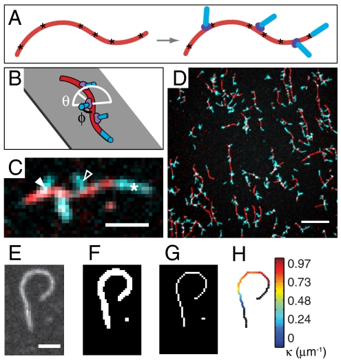
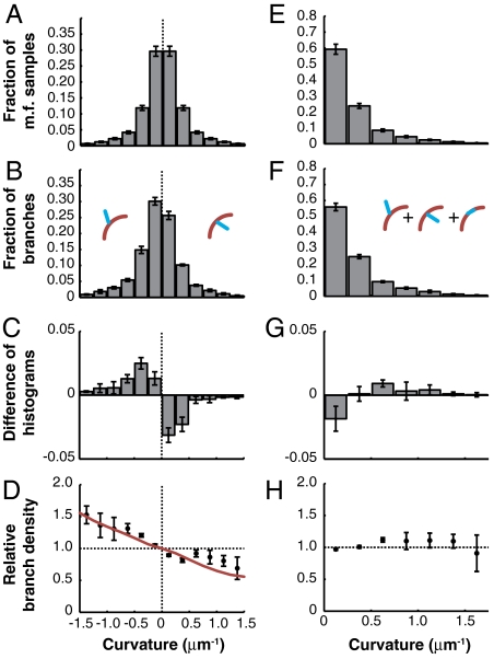
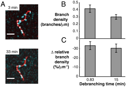
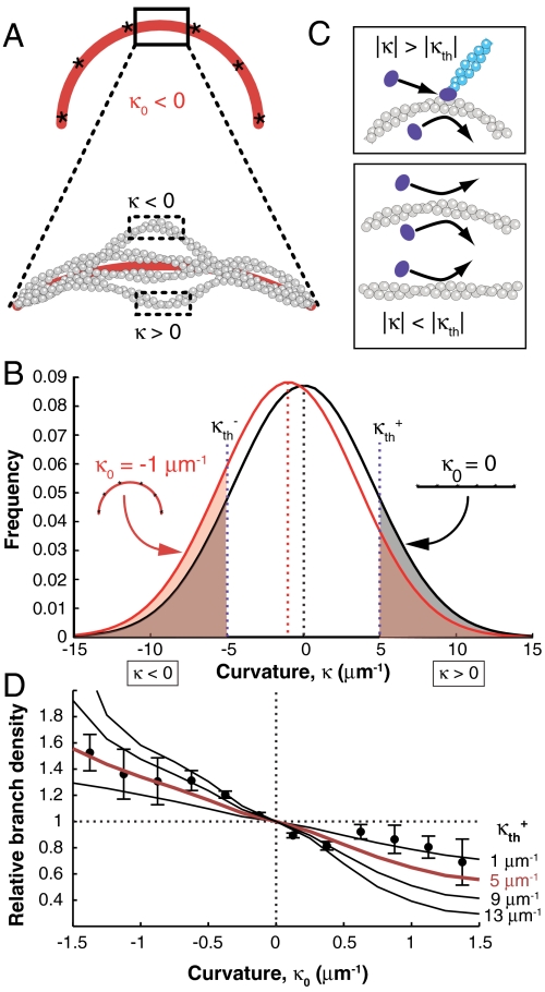
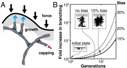
Mechanical cues affect many important biological processes in metazoan cells, such as migration, proliferation, and differentiation. Such cues are thought to be detected by specialized mechanosensing molecules linked to the cytoskeleton, an intracellular network of protein filaments that provide mechanical rigidity to the cell and drive cellular shape change. The most abundant such filament, actin, forms branched networks nucleated by the actin-related protein (Arp) 2/3 complex that support or induce membrane protrusions and display adaptive behavior in response to compressive forces. Here we show that filamentous actin serves in a mechanosensitive capacity itself, by biasing the location of actin branch nucleation in response to filament bending. Using an in vitro assay to measure branching from curved sections of immobilized actin filaments, we observed preferential branch formation by the Arp2/3 complex on the convex face of the curved filament. To explain this behavior, we propose a fluctuation gating model in which filament binding or branch nucleation by Arp2/3 occur only when a sufficiently large, transient, local curvature fluctuation causes a favorable conformational change in the filament, and we show with Monte Carlo simulations that this model can quantitatively account for our experimental data. We also show how the branching bias can reinforce actin networks in response to compressive forces. These results demonstrate how filament curvature can alter the interaction of cytoskeletal filaments with regulatory proteins, suggesting that direct mechanotransduction by actin may serve as a general mechanism for organizing the cytoskeleton in response to force.


Conflict of interest statement

The authors declare no conflict of interest.

Figures

Fig. 1.
Fig. 1.
Branching from curved filaments was observed in vitro. (A) Mother filaments (red) immobilized via biotin-streptavidin tethers (asterisks) before nucleation of branches (cyan blue) by Arp2/3 complex (violet). (B) Actin branches grow at a branch angle φ ∼ 70° to the mother filament (black line) with an azimuthal angle θ from 0° to 180° (white line). (C) Fluorescence image of actin growth at mother filament ends (white asterisk) and on branches on concave (open arrowhead) and convex (filled arrowhead) sides of mother filament curves. (Scale bar: 2 μm.) (D) Sample field of view. (Scale bar: 10 μm.) (EG) Filament image thresholded and skeletonized to an 8-connected digital curve. (Scale bar: 2 μm.) (H) Mother filament curvature measured with the tangent angle method.
Fig. 2.
Fig. 2.
Filament curvature biases branching direction. (A) Mother filament curvature distribution and (B) the distribution of mother filament curvature at branch points measured with the tangent angle method. (C) The difference and (D) the ratio of the histograms in B and A. The latter is called the relative branch density. The red curve represents the best fit (by least squares) by the fluctuation gating model with a 5 μm-1 threshold curvature. C, D, G, and H were normalized using a simulated control (see Materials and Methods). (EH) The unsigned curvature distributions corresponding to AD. Error bars: SEM, n = 5 independent experiments.
Fig. 3.
Fig. 3.
Branch stability does not affect the branching bias. (A) Actin branches (cyan blue) grown from unstabilized mother filaments (red) and incubated in buffer with unlabeled actin but without phalloidin stabilization for times shown exhibited little to no debranching when the same sample was imaged at the two time points. (Scale bars: 5 μm.). (B and C) To obtain enough images for curvature analysis, identical but separate samples were prepared with incubation times of 0.83 or 15 min. in KMEI buffer (see SI Materials and Methods) with unlabeled actin before stabilization with phalloidin and imaging. (B) We found a decrease in overall branch density between short and long incubation samples, but it was not statistically significant (p = 0.12, Welch’s t test, n = 4). (C) There was no significant difference in the slope of relative branch density with respect to curvature (Table S3, p = 0.66). Error bars: SEM.
Fig. 4.
Fig. 4.
The branching bias can be explained by a shift in the curvature fluctuations of a WLC filament tethered to a curved path. (A) Schematic of the WLC polymer tethered at six points (asterisks) to a curve with imposed curvature κ0 < 0. Fluctuations with local curvature κ < 0 and κ > 0 are possible. Curvature was calculated from the section between the middle two tethered particles, in order to avoid end effects (SI Materials and Methods). (B) Distribution of local curvature fluctuations for a filament tethered to a straight (black) or curved (red) path. Shaded areas indicate probability of branching. (C) The fluctuation gating model predicts a threshold convex local curvature beyond which stable binding and branching by the Arp2/3 complex (violet) can occur. (D) Relative branch density calculated from the ratio of the red- and black-shaded areas in B for several values of κth plotted with experimental data (also shown in Fig. 2D, with the red line corresponding to the same value of κth). Error bars: SEM.
Fig. 5.
Fig. 5.
A bias in the direction of branching can increase the total amount of actin in a branched network. (A) In a branched network, compressive forces bend filaments away from the membrane (black). Excess branching on the convex side of a bent filament creates more branches pointing toward the membrane, increasing the number of filaments pushing against the membrane (cyan blue arrows). Capping (red) can occur anywhere, but filaments can only branch in the branching zone (gray). (B) Results of a stochastic branching simulation (Materials and Methods) in which rigid branches with angles of ± 36° and -108° grow with a given bias (right column) toward the membrane. (Insets) Schematic snapshots of branching with 0% and 15% bias (gray, branching zone).

Comment in

Similar articles

Cited by

References

    1. Farge E. Mechanotransduction in development. Curr Top Dev Biol. 2011;95:243–265. - PubMed
    1. Vogel V, Sheetz M. Local force and geometry sensing regulate cell functions. Nat Rev Mol Cell Biol. 2006;7:265–275. - PubMed
    1. Hoffman BD, Grashoff C, Schwartz MA. Dynamic molecular processes mediate cellular mechanotransduction. Nature. 2011;475:316–323. - PMC - PubMed
    1. Janmey PA, McCulloch CA. Cell mechanics: Integrating cell responses to mechanical stimuli. Annu Rev Biomed Eng. 2007;9:1–34. - PubMed
    1. Fletcher DA, Mullins RD. Cell mechanics and the cytoskeleton. Nature. 2010;463:485–492. - PMC - PubMed

Publication types