. 2012 Feb 14;109(7):2325-9.
doi: 10.1073/pnas.1114740109. Epub 2012 Feb 2.

High-resolution structure of a BRICHOS domain and its implications for anti-amyloid chaperone activity on lung surfactant protein C

Affiliations

High-resolution structure of a BRICHOS domain and its implications for anti-amyloid chaperone activity on lung surfactant protein C

Hanna Willander et al. Proc Natl Acad Sci U S A. .

Abstract

BRICHOS domains are encoded in > 30 human genes, which are associated with cancer, neurodegeneration, and interstitial lung disease (ILD). The BRICHOS domain from lung surfactant protein C proprotein (proSP-C) is required for membrane insertion of SP-C and has anti-amyloid activity in vitro. Here, we report the 2.1 Å crystal structure of the human proSP-C BRICHOS domain, which, together with molecular dynamics simulations and hydrogen-deuterium exchange mass spectrometry, reveals how BRICHOS domains may mediate chaperone activity. Observation of amyloid deposits composed of mature SP-C in lung tissue samples from ILD patients with mutations in the BRICHOS domain or in its peptide-binding linker region supports the in vivo relevance of the proposed mechanism. The results indicate that ILD mutations interfering with proSP-C BRICHOS activity cause amyloid disease secondary to intramolecular chaperone malfunction.


Conflict of interest statement

The authors declare no conflict of interest.

Figures
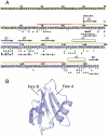
Fig. 1.
Fig. 1.
ProSP-C sequence and 3D structure of its BRICHOS domain. (A) Sequence of full-length human proSP-C. The N-terminal, situated in the cytosol, is presented in yellow. The TM and mature SP-C parts are in green. The C-terminal part of proSP-C (CTC) is shown in gray for the linker and blue for the BRICHOS domain. HDX rate constants in CTC are shown as colored lines above the sequence where red is fast, yellow is intermediate and blue is slow exchange. Secondary structure elements are shown as rectangles (helices) and arrows (β-strands). Starting position of the BRICHOS domain is labeled. Green dots, below the sequence, represent strictly conserved residues. Asterisks mark ILD mutations; the black are point mutations, the highlighted red correspond to the Δ91-93 deletion, the highlighted yellow are frameshift mutations, the two red asterisks correspond to start and end points of the Δexon 4 deletion, and the unfilled asterisk corresponds to an 18 base pair insertion. Residues in the trimer interface are labeled with black triangles. Open and filled circles identify residues on face A and B of the β-sheet, respectively. (B) Ribbon diagram representation of one subunit, with secondary structure elements β1-β2-β3-β4-α1-α2-β5 labeled. A dashed line indicates the missing region between helices α1 and α2.
Fig. 2.
Fig. 2.
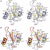
Structural conservation and location of ILD-associated mutations in the BRICHOS domain of human proSP-C. (A) Stereo view showing the BRICHOS domain as a cartoon with conserved residues as sticks. (B) As (A) but showing the targets for ILD-associated mutations. Point mutations are shown as sticks labeled with the proSP-C residue number and residue type, the Δexon4 and Δ91-93 deletion mutations are shown in red, frame shift mutations are colored yellow and identified by residue number.
Fig. 3.
Fig. 3.
Conformational changes after MD simulations. The two structures after MD simulations are superimposed on the starting X-ray structure (green). The D105N mutant in blue remains unchanged compared to the distorted WT monomer in magenta.
Fig. 4.
Fig. 4.
The linker region stabilizes substrate peptides bound to BRICHOS. HDX-MS spectra show that the presence of V7 induces significant protection from deuterium labeling in the VLEM fragment from the N-terminal linker region (top left). Similarly, a subpopulation of the V7 peptide is significantly protected from exchange when CTC is present (bottom right). The schematic model (bottom left) shows how bound target peptides can interact with the linker to form a β-hairpin, see text for details.
Fig. 5.
Fig. 5.
Amyloid in lung tissue. (A) The amyloid was strongly stained with Congo red and showed a bright green birefringence in polarized light (arrows), diagnostic of amyloid. (B) An amyloid deposit, labeled with an antibody against mature SP-C, visualized with 2,2′-diamino benzidine (brown) and then stained with Congo red and examined in polarized light. Staining with Congo red is evident in the periphery of the deposit (arrow). (C) Small amyloid deposits close to a vessel immunolabeled for SAP and in addition stained with Congo red for visualization of amyloid. Congo red staining and SAP labeling colocalize (black arrows) but SAP is also present in elastic structures (green arrow). (D) Same material as in (C), but visualized between crossed polars. [Scale bars (A, C, and D), 50 μm and (B) 20 μm.]

References

    1. Mulugeta S, Beers MF. Surfactant protein C: its unique properties and emerging immunomodulatory role in the lung. Microbes Infect. 2006;8:2317–2323. - PubMed
    1. Beers MF, Kim CY, Dodia C, Fisher AB. Localization, synthesis, and processing of surfactant protein SP-C in rat lung analyzed by epitope-specific antipeptide antibodies. J Biol Chem. 1994;269:20318–20328. - PubMed
    1. Johansson J, Szyperski T, Curstedt T, Wüthrich K. The NMR structure of the pulmonary surfactant-associated polypeptide SP-C in an apolar solvent contains a valyl-rich α-helix. Biochemistry. 1994;33:6015–6023. - PubMed
    1. Mingarro I, Nilsson I, Whitley P, von Heijne G. Different conformations of nascent polypeptides during translocation across the ER membrane. BMC Cell Biol. 2000;1 Available at http://www.biomedcentral.com/1471-2121/1/3. - PMC - PubMed
    1. Kallberg Y, Gustafsson M, Persson B, Thyberg J, Johansson J. Prediction of amyloid fibril-forming proteins. J Biol Chem. 2001;276:12945–12950. - PubMed

Publication types

MeSH terms

Substances

Associated data

LinkOut - more resources