. 2012 Feb 7;109(6):E336-42.
doi: 10.1073/pnas.1114965109. Epub 2012 Jan 17.

CD4 and CD8 T cells require different membrane gangliosides for activation

Affiliations

CD4 and CD8 T cells require different membrane gangliosides for activation

Masakazu Nagafuku et al. Proc Natl Acad Sci U S A. .

Abstract

Initial events of T-cell activation involve movement of the T-cell receptor into lipid rafts. Gangliosides are major components of lipid rafts. While investigating T-cell activation in ganglioside-deficient mice, we observed that CD4(+) and CD8(+) T cells required different ganglioside subsets for activation. Activation of CD4(+) T cells from GM3 synthase-null mice, deficient in GM3-derived gangliosides, is severely compromised, whereas CD8(+) T-cell activation is normal. Conversely, in cells from GM2/GD2 synthase-null mice, expressing only GM3 and GD3, CD4(+) T-cell activation is normal, whereas CD8(+) T-cell activation is deficient. Supplementing the cells with the corresponding missing gangliosides restores normal activation. GM3 synthase-null mice do not develop experimental asthma. Distinct expression patterns of ganglioside species in CD4(+) T and CD8(+) T cells, perhaps in uniquely functional lipid rafts, define immune functions in each T-cell subset. Control of ganglioside expression would offer a strategy targeting for specific T-cell subpopulations to treat immune diseases.


Conflict of interest statement

The authors declare no conflict of interest.

Figures
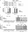
Fig. 1.
Fig. 1.
Distinct selectivity of ganglioside species for CD4+ T and CD8+ T activation. (A) Ganglio-series glycosphingolipids are synthesized from ceramide and are divided into o-, a-, and b-series species. (B) Activation of CD4+ T and CD8+ T cells upon TCR-mediated stimulation. Purified peripheral CD4+ T and CD8+ T cells from WT, GM3S null, and GM2/GD2S null mice (20–25 mice each) were left unstimulated (NS) or were stimulated for 72 h with anti-CD3 antibody plus anti-CD28 antibody or with PMA plus ionomycin (Io). Proliferative responses were determined after an 8-h pulse with XTT reagent. One representative of three experiments is presented. *P < 0.01. (C and D) Functional rescue experiments by supplementation of gangliosides. Purified peripheral CD4+ T (C) and CD8+ T (D) cells from WT, GM3S null, and GM2/GD2S null mice were pretreated for 2 h with the indicated GSL (5 μg/mL). The cells were left unstimulated (NS) or were stimulated for 72 h with anti-CD3 antibody plus anti-CD28 antibody. Proliferative responses were determined after an 8-h pulse with XTT reagent. Data are representative of more than three experiments.
Fig. 2.
Fig. 2.
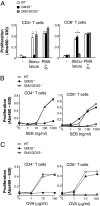
Antigen-specific responses of CD4+ T and CD8+ T cells of GM3S null and GM2/GD2S null mice. (A and B) Peripheral CD4+ T and CD8+ T cells were purified from WT, GM3S null, and GM2/GD2S null mice. (A) Mixed lymphocyte reaction. The cells were left untreated (−), mixed with MMC-treated MHC-mismatched splenocytes from BALB/c as stimulators, or treated with PMA plus ionomycin (Io) for 72 h. (B) Superantigen-induced activation. The indicated cells were stimulated with indicated amounts of Staphylococcal enterotoxin B (SEB) and MMC-treated WT splenocytes as APCs. (C) Antigen-specific T-cell responses. Purified CD4+T and CD8+T cells from WT, GM3S null and GM2/GD2S null mice were obtained 7 d after immunization with TNP-OVA. The resulting cells were cocultured with MMC-treated WT splenocytes as APCs, together with the indicated amounts of OVA peptide. Proliferative responses were determined after an 8-h pulse with XTT reagent. Data are representative of three experiments. *P < 0.01 vs. WT.
Fig. 3.
Fig. 3.
Glycosphinogolipid expression patterns in thymocytes and peripheral primary CD4+ T and CD8+ T cells in WT, GM3S−/−, and GM2/GD2S−/− mice. Neutral and acidic lipids were obtained from thymocytes, CD4+ T cells, and CD8+ T cells of WT, GM3S−/− (G3), and GM2/GD2S−/− (G2) mice. (A) The acidic lipids were separated on HPTLC plates and were stained with HRP-conjugated choleragenoid (CTx-B). An arrow indicates the origin for TLC. White and black arrowheads indicate GM1a and extended-GM1b bands, respectively. (B and C) Acidic (B) and neutral (C) lipids were separated on HPTLC plates and visualized with orcinol-sulfuric acid. White and black arrowheads in (B) indicate GM3 and GD3 bands, respectively. Fuc-GM1a, fucosylated GM1a; std, standard lipids.
Fig. 4.
Fig. 4.
Distinct expression of ganglioside species during T-cell repertoire selection. (A) Gene expression of GM3S and GM2/GD2S was determined by quantitative real-time PCR analysis. Data represent the relative gene expression in peripheral T-cell subsets compared with that in thymocytes (set to 1.0 separately for each target gene). *P < 0.05. (B) Gangliosides of mouse thymocytes and CD4+ and CD8+ T cells analyzed by LC-MS. Peak areas of GM1, GD1, GalNAcGM1b, and extended-GM1b were determined in the mass chromatograms obtained by C30 column chromatography-MS with GM3 (NeuAc, d18:1–14:0) as an internal standard. GM1 includes GM1a [N-glycolylneuraminic acid (NeuGc)] and GM1b (NeuGc), each carrying d18:1–16:0, –18:0, –20:0, –22:0, –24:1, and –24:0. GD1 includes GD1b (NeuGc) and GD1c (NeuGc) each carrying d18:1–16:0, –18:0, –20:0, –22:0, –24:1, and –24:0. GD1a was not detectable in any of the three cell types.
Fig. 5.
Fig. 5.
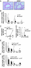
Allergic airway inflammation in GM3S null mice. (A and B) Sensitized WT and GM3S null mice were challenged with OVA. Five days after the challenge, BAL fluid and lung tissues were collected. The representative lung sections stained with PAS (A) and the numbers of total cells (Total), macrophages (Mφ), eosinophils (Eo), neutrophils (Neu), and lymphocytes (Lym) in BAL fluids (B) are shown. (WT, n = 7; GM3S null, n = 9). (Scale bar in A, 200 μm.) (C) Sera from sensitized WT and GM3S null mice were obtained at 1 (WT, n = 6; GM3S null, n = 6), 3 (WT, n = 6; GM3S null, n = 6) and 5 d (WT, n = 6; GM3S null, n = 6) after OVA challenge. OVA-specific IgE in the sera was measured. (D) Cytokine levels in BAL fluid (WT, n = 6; GM3S null, n = 6) were measured 1 d after challenge. (E and F) Cell numbers in BAL fluid of mice adoptively transferred with CD4+ T cells. CD4+ T cells from sensitized WT and GM3S null mice (E) or GM2/GD2S null mice (F) were transferred to naive WT mice. BAL fluid was obtained from the recipient mice 5 d after OVA challenge [(E) WT recipient, n = 6; GM3S null recipient, n = 7; (F) WT recipient, n = 10; GM2/GD2S null recipient, n = 6]. The numbers of total cells (Total), macrophages (Mφ), eosinophils (Eo), neutrophils (Neu), and lymphocytes (Lym) in BAL fluid were counted. *P < 0.05, **P < 0.01.
Fig. P1.
Fig. P1.
Selection of T cells to be produced (repertoire selection) by ganglioside selection. (A) The core biosynthetic pathway of gangliosides. Staining by CTx-B detects not only GM1a ganglioside but also extended-GM1b. (B) Distinct difference of lipid rafts in individual T-cell subsets. The repertoire selection from immature thymocytes to mature T-cell subpopulations is accompanied by selective ganglioside expression. Analysis confirms that CD4+ T cells dominantly express a-series gangliosides due to up-regulation (increased expression) of GM3S genes. Meanwhile, CD8+ T cells carry o-series gangliosides due to down-regulation (decreased expression) of GM3S genes and up-regulation of GM2/GD2S expression. This result suggests that each T-cell subset has unique rafts composed of different ganglioside types and that these rafts provide distinct functions following stimulation of T cells via their receptors (TCRs). This ganglioside selection process may be indispensable in the formation of distinct and functional lipid rafts in mature T cells.

References

    1. Simons K, Ikonen E. Functional rafts in cell membranes. Nature. 1997;387:569–572. - PubMed
    1. Dykstra M, Cherukuri A, Sohn HW, Tzeng SJ, Pierce SK. Location is everything: Lipid rafts and immune cell signaling. Annu Rev Immunol. 2003;21:457–481. - PubMed
    1. Harder T, Rentero C, Zech T, Gaus K. Plasma membrane segregation during T cell activation: Probing the order of domains. Curr Opin Immunol. 2007;19:470–475. - PubMed
    1. Kovacs B, et al. Human CD8+ T cells do not require the polarization of lipid rafts for activation and proliferation. Proc Natl Acad Sci USA. 2002;99:15006–15011. - PMC - PubMed
    1. Gómez-Móuton C, et al. Segregation of leading-edge and uropod components into specific lipid rafts during T cell polarization. Proc Natl Acad Sci USA. 2001;98:9642–9647. - PMC - PubMed

Publication types

MeSH terms

LinkOut - more resources