. 2012 Feb 14;109(7):E432-41.
doi: 10.1073/pnas.1115346109. Epub 2012 Jan 17.

Ancestral capture of syncytin-Car1, a fusogenic endogenous retroviral envelope gene involved in placentation and conserved in Carnivora

Affiliations

Ancestral capture of syncytin-Car1, a fusogenic endogenous retroviral envelope gene involved in placentation and conserved in Carnivora

Guillaume Cornelis et al. Proc Natl Acad Sci U S A. .

Abstract

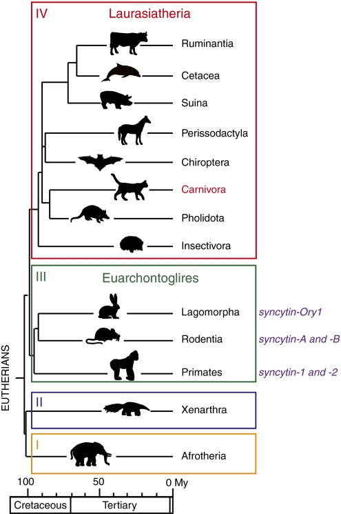
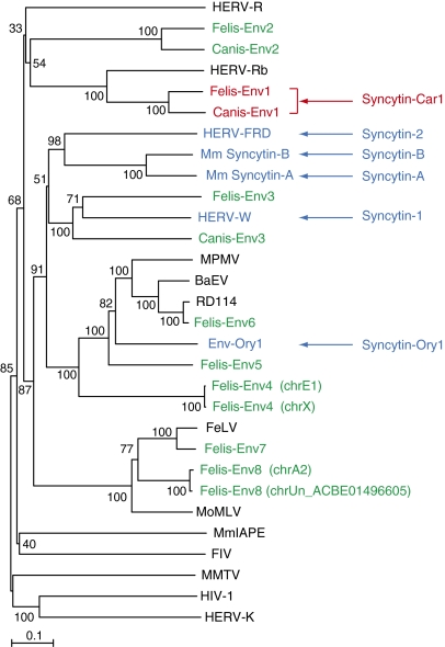
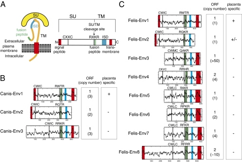
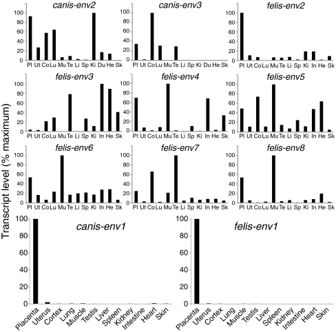
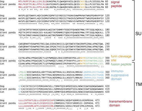
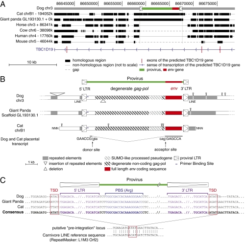
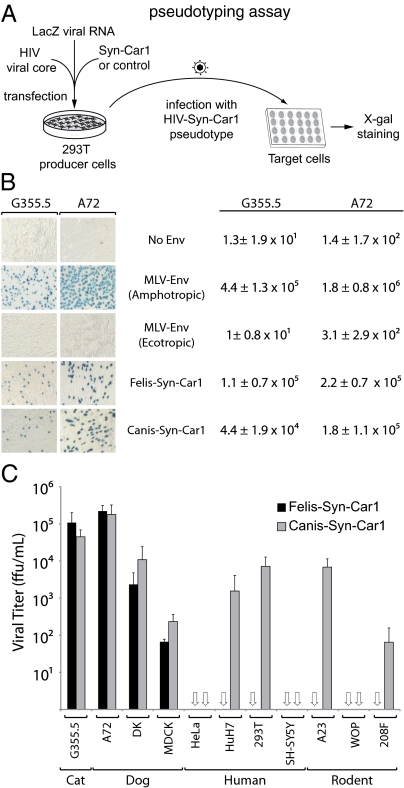
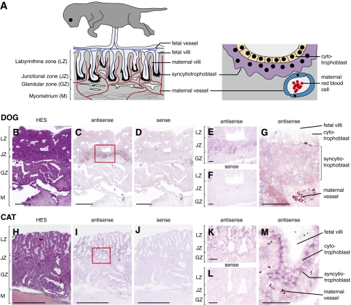
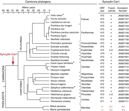
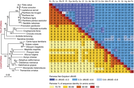
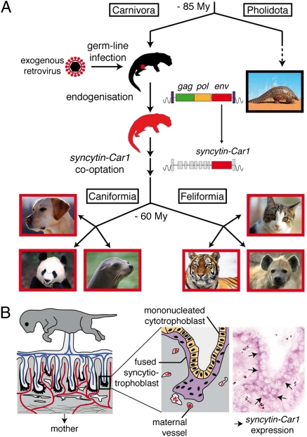
Syncytins are envelope protein genes of retroviral origin that have been captured for a function in placentation. Two such genes have already been identified in simians, two distinct, unrelated genes have been identified in Muridae, and a fifth gene has been identified in the rabbit. Here, we searched for similar genes in the Laurasiatheria clade, which diverged from Euarchontoglires--primates, rodents, and lagomorphs--shortly after mammalian radiation (100 Mya). In silico search for envelope protein genes with full-coding capacity within the dog and cat genomes identified several candidate genes, with one common to both species that displayed placenta-specific expression, which was revealed by RT-PCR analysis of a large panel of tissues. This gene belongs to a degenerate endogenous retroviral element, with precise proviral integration at a site common to dog and cat. Cloning of the gene for an ex vivo pseudotype assay showed fusogenicity on both dog and cat cells. In situ hybridization on placenta sections from both species showed specific expression at the level of the invasive fetal villi within the placental junctional zone, where trophoblast cells fuse into a syncytiotrophoblast layer to form the maternofetal interface. Finally, we show that the gene is conserved among a series of 26 Carnivora representatives, with evidence for purifying selection and conservation of fusogenic activity. The gene is not found in the Pholidota order and, therefore, it was captured before Carnivora radiation, between 60 and 85 Mya. This gene is the oldest syncytin gene identified to date, and it is the first in a new major clade of eutherian mammals.


Conflict of interest statement

The authors declare no conflict of interest.

Figures

Fig. 1.
Fig. 1.
Phylogeny of Eutherians and previously identified syncytin genes. Eutherians can be grouped into four major clades: Afrotheria (I), Xenarthra (II), Euarchontoglires (III), and Laurasiatheria (IV). Adapted from ref. . Branch length is proportional to time (in million years), and the orders where syncytins have been identified to date are indicated with the corresponding gene name.
Fig. 2.
Fig. 2.
Retroviral envelope protein-based phylogenetic tree with the identified Canis- and Felis-Env protein candidates. The tree was determined by the neighbor-joining method using envelope amino acid sequences from murine and human endogenous retroviruses and a series of infectious retroviruses. The horizontal branch length is proportional to the percentage of amino acid substitutions from the node (scale bar on the left), and the percent bootstrap values obtained from 1,000 replicates are indicated at the nodes. The two pairs of Felis-Env proteins that were grouped into single families of elements (Felis-Env4 and Felis-Env8) are distinguished by indication of their chromosomal position. BaEV, baboon endogenous virus; FeLV, feline leukemia virus; FIV, feline immunodeficiency virus; HERV, human ERV; MoMLV, moloney murine leukemia virus; MmIAPE, Mus musculus intracisternal A-type particle with an envelope gene; MMTV, murine mammary tumor virus; MPMV, Mason–Pfizer monkey virus; RD114, feline endogenous type-C retrovirus.
Fig. 3.
Fig. 3.
Structure of a canonical retroviral envelope protein and characterization of the identified dog and cat candidates. (A) Schematic representation of a retroviral envelope protein with the SU and TM subunits delineated and the furin cleavage site (consensus: R/K-X-R/K-R) between the two subunits together with the C-X-X-C domain involved in SU–TM interaction indicated; the hydrophobic signal peptide (red), fusion peptide (green), transmembrane domain (red), and putative immunosuppressive domain (ISD; blue) are also indicated. (B and C) Characterization of the dog and cat candidate envelope proteins, respectively. (Left) The hydrophobicity profile for each candidate is shown with the canonical structural features listed in A positioned when present (same color code). (Right) Number of full-length coding sequences (ORF) of env genes within each family of element and total number of genomic copies (in parenthesis) together with indication for specific placental expression (details in Fig. 4).
Fig. 4.
Fig. 4.
Real-time qRT-PCR analysis of the candidate env gene transcripts from dog and cat. Transcript levels are expressed as percent of maximum and were normalized relative to the amount of a control gene (PPIA) (Methods) RNA. The two placenta-specific canis-env1 and felis-env1 genes are displayed in enlarged panels in the fourth row; the same tissues (abbreviated and displayed in the same order) for the nine other env gene candidates are shown. Values for the placenta and uterus are the means of at least five samples for both dog and cat; RNAs of other tissues are from Zyagen.
Fig. 5.
Fig. 5.
Primary sequences and alignment of Canis-Env1, Felis-Env1, and Ailur-Env1. Primary amino acid sequence and characteristic structural features of the homologous Env protein from dog, cat, and giant panda. Same color code and abbreviations as in Fig. 3; asterisks indicate amino acid identity, and colons indicate amino acid similarity.
Fig. 6.
Fig. 6.
Characterization of the dog, cat, and giant panda env1-containing endogenous retrovirus and its integration site. (A) Evidence for orthology of the dog, cat, and panda sequences. The canis-env1–containing provirus (shown in green with its LTRs schematized by boxed triangles and the env gene shown in red) was used as a reference, and synteny between the dog, cat, giant panda, horse, cow, human, and mouse genomes was determined with the Comparative Genomic tool of the UCSC Genome Browser (http://genome.ucsc.edu/); the position of exons (vertical red lines) of the resident TBC1D19 gene and the sense of transcription (blue arrows) are indicated. Homologous regions are shown as black boxes, nonhomologous regions are shown as thin lines (not to scale), and gaps are shown as dotted lines. (B) Structure of the corresponding proviruses and integration sites. Homologous regions common to all three sequences are aligned, with the insertions and deletions represented by triangles placed above and below each sequence, respectively; repeated mobile elements (dark gray) as identified by the RepeatMasker website program are positioned. The proviral LTRs, the degenerate gag-pol gene, and the env gene coding sequence are indicated together with the SUMO-like processed pseudogene inserted within the provirus (the symbols used are given below the panel). Splice sites for the env subgenomic transcript as determined by RT-PCR from dog and cat placental RNA are indicated together with the donor and acceptor site sequences. (C) Sequence alignment of proviral and flanking sequences: evidence for retroviral integration. Characteristic sequences are shown, including LTRs (purple with TG … CA borders), primer binding site (blue) for an arginine tRNA, and target site duplication (red boxes). Sequence alignment of the reconstituted, putative preintegration site with the Carnivore LINE L1M3 Orf2 (prototypic reference sequence from the RepeatMasker website) provides evidence for precise proviral integration into a preexisting ancestral LINE.
Fig. 7.
Fig. 7.
Syncytin-Car1 is a fusogenic retroviral envelope protein. (A) Schematic representation of the assay for cell infection with Syncytin-Car1–pseudotyped virus particles. Pseudotypes are produced by cotransfection of human 293T cells, with expression vectors for the HIV-1 core, Syncytin-Car1 proteins (or control retroviral envelope proteins or an empty vector), and β-Gal encoded by a LacZ-containing retroviral transcript. Productive cell supernatants are then assayed for infection of the indicated target cells, which are X-gal–stained 3 d postinfection. Syn-Car1: Syncytin-Car1. (B) X-gal–stained target cells (Left) and viral titers (Right) for particles pseudotyped with Felis-Syn-Car1, Canis-Syn-Car1, or control Env proteins from murine leukemia viruses of either the ecotropic (infecting only murine cells) or the amphotropic (infecting both murine and nonmurine cells) subtype. Target cells were cat (G355.5) and dog (A72) cells. Values (focus forming unit per milliliter ± SD) are the means from three independent experiments. (C) Viral titers for particles pseudotyped with Felis-Syn-Car1 or Canis-Syn-Car1 assayed on a panel of target cells from cat (G355.5), dog (A72, MDCK, and DK), human (HeLa, HuH7, 293T, and SH-SY5Y), or rodent (A23, WOP, and 208F). Titers are corrected for the background values of control particles without an envelope protein, and they are the means from at least two independent experiments (focus forming unit per milliliter ± SD; open arrows indicate null values).
Fig. 8.
Fig. 8.
Structure of the Carnivora placenta and in situ hybridization for syncytin-Car1 expression on dog and cat placental sections. (A) Schematic representation of the Carnivora placenta (A Left) with, from mother to fetus, the myometrium (M), the glandular zone (GZ), the junctional zone (JZ), and the labyrinthine zone (LZ). The maternal and fetal vessels are schematized, with the invasive syncytiotrophoblast at the fetomaternal interface colored in black at the tip of the invading fetal villi. An enlarged view (A Right) shows the multinucleated syncytiotrophoblast (purple) generated by fusion from the underlying mononucleated cytotrophoblast cells (yellow) forming the interface between the fetal compartments and the maternal vessels (with the red blood cells schematized). Sections of placenta from dog (B–G) and cat (H–M) at midgestation. (B and H) Hematoxylin–eosin–saffron staining (HES) of the placental section with the four layers indicated. (C–G and I–M) In situ hybridization on serial sections observed at different magnifications using digoxigenin-labeled antisense (C, E, G, I, K, and M) or sense (negative control; D, F, J, and L) riboprobes revealed with an alkaline phosphatase-conjugated antidigoxigenin antibody. (E, F, K, and L) Intermediate magnification of the junctional domain delineated in C and I (red boxed areas). (G and M) Higher magnification with the fetal villi, the labeled syncytiotrophoblast, and the maternal vessels clearly visible. (Scale bars: BD and HJ, 1 mm; E, F, K, and L, 100 μm; G and M, 50 μm.)
Fig. 9.
Fig. 9.
Entry date and conservation of syncytin-Car1 in the Carnivora radiation. (Left) Carnivora phylogenetic tree with the Pholidota outgroup indicated. Adapted from refs. , , and . Horizontal branch length is proportional to time (scale bar at the top). The names of the 26 Carnivora species (from both the Feliformia and Caniformia suborders) tested for the presence of the syncytin-Car1 gene are indicated together with the names of their corresponding families. (Right) The length (in amino acids) of the Syncytin-Car1 proteins that were identified for each species and the accession number for the sequences that were deposited in GenBank are indicated; the fusogenic activity for each cloned gene, as determined by the pseudotyping assay described in Fig. 7, is provided (n.d., not determined). For the two Manidae tested (from the Pholidota Order), syncytin-Car1 was not detected (n.r., not relevant), thus dating back acquisition of the gene between 60 and 85 Mya. Asterisks indicate species where placenta-specific expression could be shown (Figs. 4 and 8 for dog [C. lupus familiaris] and cat [F. catus], and Fig. S1 for sea lion [Zalophus californianus]).
Fig. 10.
Fig. 10.
Sequence conservation and evidence for purifying selection of syncytin-Car1 in Carnivora. (Left) Syncytin-Car1–based phylogenetic tree determined using amino acid alignment of the Syncytin-Car1 proteins identified in Fig. 9 by the neighbor-joining method. The horizontal branch length and scale indicate the percentage of amino acid substitutions. Percent bootstrap values obtained from 1,000 replicates are indicated at the nodes. (Right) Double-entry table for (lower triangle) the pairwise percentage of amino acid sequence identity between the syncytin-Car1 gene among the indicated species and (upper triangle) the pairwise Nei–Gojobori (30) dN/dS; dN/dS between Panthera leo and P. leo krugeri could not be determined (only one nonsynonymous and no synonymous mutation between both species; n.r. not relevant). A color code is provided below the table for both series of values.
Fig. P1.
Fig. P1.
From infectious retroviruses to captured syncytin genes with a placental function. (A) Gene capture through infectious retrovirus endogenization. Infection of a Carnivora ancestor taking place after the Carnivora/Pholidota split 85 Mya, with viral integration into the genome; in the absence of selective pressure, the retroviral gag and pol genes have lost their coding capacity (genetic drift), whereas cooptation of the retroviral env gene resulted in the maintenance of its coding capacity in the course of Carnivora evolution (with the major Caniformia/Feliformia split taking place 60 Mya). (B) Specific placental expression of the captured syncytin-Car1 gene within the fetomaternal interface at the level of the fused syncytiotrophoblast layer. The placenta with the maternal and fetal blood vessels is schematized together with the fetomaternal interface, where cells fuse into the syncytiotrophoblast as a result of syncytin-Car1 expression that is revealed by in situ hybridization.

Comment in

Similar articles

Cited by

References

    1. Mi S, et al. Syncytin is a captive retroviral envelope protein involved in human placental morphogenesis. Nature. 2000;403:785–789. - PubMed
    1. Blond JL, et al. An envelope glycoprotein of the human endogenous retrovirus HERV-W is expressed in the human placenta and fuses cells expressing the type D mammalian retrovirus receptor. J Virol. 2000;74:3321–3329. - PMC - PubMed
    1. Mallet F, et al. The endogenous retroviral locus ERVWE1 is a bona fide gene involved in hominoid placental physiology. Proc Natl Acad Sci USA. 2004;101:1731–1736. - PMC - PubMed
    1. Cáceres M, Thomas JW, Thomas JW. NISC Comparative Sequencing Program The gene of retroviral origin Syncytin 1 is specific to hominoids and is inactive in Old World monkeys. J Hered. 2006;97:100–106. - PubMed
    1. Blaise S, de Parseval N, Bénit L, Heidmann T. Genomewide screening for fusogenic human endogenous retrovirus envelopes identifies syncytin 2, a gene conserved on primate evolution. Proc Natl Acad Sci USA. 2003;100:13013–13018. - PMC - PubMed

Publication types

Associated data