. 2012 Feb 14;109(7):2485-90.
doi: 10.1073/pnas.1115369109. Epub 2012 Jan 30.

Memory B cells in the lung participate in protective humoral immune responses to pulmonary influenza virus reinfection

Affiliations

Memory B cells in the lung participate in protective humoral immune responses to pulmonary influenza virus reinfection

Taishi Onodera et al. Proc Natl Acad Sci U S A. .

Abstract

After pulmonary virus infection, virus-binding B cells ectopically accumulate in the lung. However, their contribution to protective immunity against reinfecting viruses remains unknown. Here, we show the phenotypes and protective functions of virus-binding memory B cells that persist in the lung following pulmonary infection with influenza virus. A fraction of virus-binding B-cell population in the lung expressed surface markers for splenic mature memory B cells (CD73, CD80, and CD273) along with CD69 and CXCR3 that are up-regulated on lung effector/memory T cells. The lung B-cell population with memory phenotype persisted for more than 5 mo after infection, and on reinfection promptly differentiated into plasma cells that produced virus-neutralizing antibodies locally. This production of local IgG and IgA neutralizing antibody was correlated with reduced virus spread in adapted hosts. Our data demonstrates that infected lungs harbor a memory B-cell subset with distinctive phenotype and ability to provide protection against pulmonary virus reinfection.


Conflict of interest statement

The authors declare no conflict of interest.

Figures
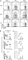
Fig. 1.
Fig. 1.
Kinetics and surface phenotypes of HA-binding IgM/D B-cell populations. (A) Cells recovered at the indicated time points after infection were subjected to flow-cytometric analysis (n = 4–5). Representative flow data for HA-binding/CD38 expression by IgM/Ddump B cells (Fig. S1) are shown. (B) Absolute cell number of CD38+ and CD38 cells within the lymphocyte gate was plotted. (C) The frequencies of cells expressing CD69, CXCR3, or IgA are plotted. In B and C, each circle represents the result for an individual mouse (lung and spleen) and two to four pooled mice (MLN). **P < 0.01.
Fig. 2.
Fig. 2.
Reactivation and terminal differentiation of the memory-like B-cell population in lung following secondary infection. (A) BALB/c mice were infected twice, with an 80-d interval. Mice were treated intraperitoneally with 1 mg BrdU for 48 h before analysis, and cells from the indicated organs were subjected to intracellular analysis for BrdU. Representative flow data for BrdU staining of HA-binding IgM/DCD38+ B cells are presented (n = 3). (B) HA-binding/CD138 expression among B220dulldump cells and IgA expression among HA-binding CD138+ cells are presented (n = 3–5).
Fig. 3.
Fig. 3.
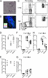
Protective function of the memory-like B-cell population against secondary infection. (A) CB17-scid mice were injected intranasally with CpG and i.v. with BALB/c splenocytes. Ten days after treatment, lung sections were stained with HE (A) or B220 (green), CD90 (red), and DAPI (blue) (B). A representative section (n = 6) is presented. Arrowheads indicate iBALT-like structures. (Scale bar: 500 μm in A and 100 μm in B). (C) HA-binding IgM/DCD38+ B cells were sorted from pooled lungs and spleens (n = 10). (D and E) CpG-treated (D) or untreated (E) CB17-scid mice were reconstituted with either lung or splenic memory-like B-cell population (3,000 cells per mouse) together with splenic B cells from naïve mice and CD4+ T cells from infected mice. Splenic naïve B cells were added to prevent the loss of small numbers of memory-like B-cell population after sorting. On day 6 postinfection, virus titers in BALFs were determined. (F and G) HA-binding IgG and IgA-secreting plasma cells in the lungs of CpG-treated (F) or untreated (G) mice were enumerated by ELISPOT. (H) HA-binding IgG and IgA Ab titers in BALFs of CpG-treated mice were estimated by ELISA. In DH, each circle represents the result for an individual mouse. *P < 0.05; **P < 0.01.
Fig. 4.
Fig. 4.
Relative contribution of IgG and IgA Abs in BALFs to virus-neutralization. (A) Seven days after secondary challenge of BALB/c mice, anti-HA IgG and IgA Ab titers in BALFs were detected by ELISA. Each line represents the result for an individual mouse. (B) Virus-neutralization Ab titers in BALFs were determined by in vitro assay using MDCK cells. In B, each circle represents the result for an individual mouse. *P < 0.05; **P < 0.01.
Fig. 5.
Fig. 5.
Virus-dependent recruitment of IgA-secreting plasma cells from IgA memory B cells. (A) CB17-scid mice were reconstituted similarly to Fig. 3D and boosted with inactivated viruses. At day 7 after boosting, the numbers of HA-binding IgG+ and IgA+ plasma cells were enumerated and plotted. (B) The frequencies of IgA+ plasma cells among IgG+/IgA+ plasma cells are shown. (C) IgA+ and IgA memory B cells were sorted from pooled lung cells and splenocytes (n = 10). (D) The recipient mice were reconstituted with 200 IgA+ or IgA memory B cells in the lungs together with B cells and CD4+ T cells, and the numbers of IgG+ and IgA+ plasma cells were determined after boosting. (E) Mice were reconstituted with IgA memory B cells in the lungs, and the numbers of IgG+ and IgA+ plasma cells were determined after boosting with inactivated viruses or rHA. In A, B, D, and E, each circle represents the result for an individual recipient mouse. *P < 0.05; **P < 0.01.

References

    1. Dörner T, Radbruch A. Antibodies and B cell memory in viral immunity. Immunity. 2007;27:384–392. - PubMed
    1. Sallusto F, Lanzavecchia A, Araki K, Ahmed R. From vaccines to memory and back. Immunity. 2010;33:451–463. - PMC - PubMed
    1. Coro ES, Chang WL, Baumgarth N. Type I IFN receptor signals directly stimulate local B cells early following influenza virus infection. J Immunol. 2006;176:4343–4351. - PubMed
    1. Moyron-Quiroz JE, et al. Role of inducible bronchus associated lymphoid tissue (iBALT) in respiratory immunity. Nat Med. 2004;10:927–934. - PubMed
    1. Jones PD, Ada GL. Persistence of influenza virus-specific antibody-secreting cells and B cell memory after primary murine influenza virus infection. Cell Immunol. 1987;109:53–64. - PubMed

Publication types