. 2012 Feb 21;109(8):3077-82.
doi: 10.1073/pnas.1116107109. Epub 2012 Jan 30.

RNA virus microRNA that mimics a B-cell oncomiR

Affiliations

RNA virus microRNA that mimics a B-cell oncomiR

Rodney P Kincaid et al. Proc Natl Acad Sci U S A. .

Abstract

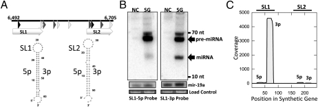
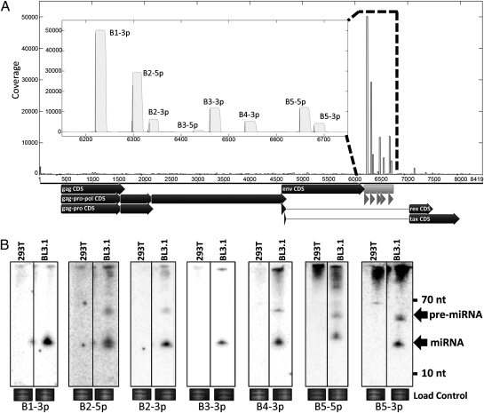
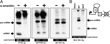
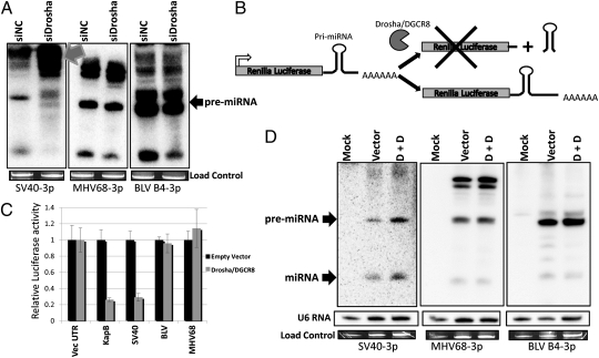
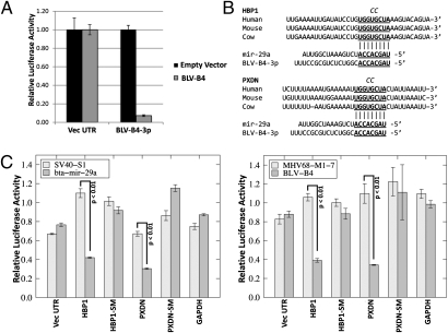
MicroRNAs (miRNAs) are small RNAs that play a regulatory role in numerous and diverse eukaryotic cellular processes. Virus-encoded miRNAs have garnered much interest, although the functions of most remain to be deciphered. To date, readily detectable, evolutionarily conserved natural miRNAs have only been identified from viruses with DNA genomes. Combined with the fact that most miRNAs are generated from endonucleolytic cleavage of longer transcripts, this finding has led to a common conception that naturally occurring RNA viruses will not encode miRNAs to avoid unproductive cleavage of their genomes or mRNAs. Here we demonstrate that the bovine leukemia virus (BLV), a retrovirus with an RNA genome, encodes a conserved cluster of miRNAs that are transcribed by RNA polymerase III (pol III). Thus, the BLV miRNAs avoid the conundrum of genome/mRNA cleavage because only the subgenomic pol III transcripts are efficiently processed into miRNAs. BLV infection is strongly associated with B-cell tumors in cattle. Because most cells in BLV-associated tumors express little viral mRNAs or proteins, exactly how BLV contributes to tumorigenesis has remained a decades-long unsolved mystery. One BLV miRNA, BLV-miR-B4, shares partial sequence identity and shared common targets with the host miRNA, miR-29. As miR-29 overexpression is associated with B-cell neoplasms that resemble BLV-associated tumors, our findings suggest a possible mechanism contributing to BLV-induced tumorigenesis.


Conflict of interest statement

The authors declare no conflict of interest.

Figures

Fig. 1.
Fig. 1.
A combined computational and synthetic approach identifies a candidate BLV-encoded miRNA. (A) Schematic of the organization of the predicted and synthesized BLV miRNA gene region. The predicted A Box-like, B Box-like, and terminator-like sequences are represented by black, gray, and white triangles respectively. Predicted stem-loop structures (SL1 and SL2) are indicated with white arrows, and Mfold RNA secondary-structure predictions for the predicted miRNA stem-loop sequences are presented below. The genomic coordinates refer to BLV reference sequence NC_001414. (B) Northern blot analysis of 293T cells transfected with the synthetic BLV miRNA gene construct (SG lanes) or negative control empty vector (NC lanes). As a load control and to provide a rough comparison of levels to a host miRNA, blots were stripped and reprobed for host mir-19a. An additional load control is ethidium bromide-stained low molecular weight RNA. (C) Small RNA profiling of 293T cells transfected with the synthetic BLV miRNA gene construct plotted as previously described (44). The vertical axis depicts small RNA read coverage observed; the horizontal axis shows the relative position within the synthetic gene sequence. Stem-loops SL1 and SL2 correspond to miRNAs BLV miR-B4 and B5 in subsequent analysis.
Fig. 2.
Fig. 2.
Small RNA profiling and Northern blots identify BLV-encoded miRNAs expressed in BL3.1 cells. (A) Small RNAs sequenced from BL3.1 cells were mapped to the BLV genome. (Inset) Detail of the newly identified miRNA-encoding region with black impulses representing start counts and gray filled area representing coverage. Below the x axis of the main graph, annotated coding sequences are displayed as black arrows. The miRNA-encoding region is indicated in light gray and individual miRNAs are represented by dark gray arrows. B4 and B5 correspond to the synthetic sequences SL1 and SL2 of Fig. 1, respectively. (B) Northern blot analysis confirms candidate miRNAs. RNA harvested from the persistently infected BL3.1 cell line or the negative control 293T cells was probed with oligonucleotides specific to the abundantly mapped sequences. The load control is ethidium bromide-stained low molecular weight RNA.
Fig. 3.
Fig. 3.
BLV miRNA B4 is transcribed by RNA Pol III. (A) Northern blot analysis of SV40-S1, MHV68-M1-7, and BLV-B4 miRNA expression vectors transfected into HEK293T cells with or without treatment of the RNA pol II inhibitor α-amanitin. The load control is ethidium bromide-stained low molecular weight RNA. (B) Northern blot analysis of RNA from 293T cells transfected with the wild-type or B Box mutant BLV-B4 miRNA expression vectors. The load control is ethidium bromide-stained low molecular weight RNA. Schematic indicates organization of the BLV miR-B4 promoter region and location of the B Box promoter element mutation.
Fig. 4.
Fig. 4.
BLV encoded miRNAs are not processed by Drosha. (A) Northern blot analysis of the SV40-S1, MHV68-M1-7, and BLV-B4 miRNA expression vectors cotransfected into HEK293T cells with either Drosha siRNA (siDrosha) or a negative control (siNC) siRNA. Gray arrow indicates accumulated unprocessed miRNA precursors. The load control is ethidium bromide-stained low molecular weight RNA. (B) Schematic of luciferase-based Drosha cleavage assay. Drosha-cleavable stem-loop structures located in the 3′UTR of the luciferase reporter result in endonucleolytic cleavage of pre-mRNA transcripts and reduced luciferase activity. (C) Renilla luciferase constructs with control pri-miRNAs or the entire BLV miRNA cluster-encoding region cloned into the 3′ UTR region were either cotransfected with empty vector or Drosha and DGCR8-expression vectors. The control Renilla reporter containing the vector-derived 3′UTR is labeled “Vec UTR.” The relative luciferase activity ratio of Renilla to firefly is graphed and normalized to the Renilla luciferase vector 3′ UTR. Error bars indicate SD of three replicates. (D) Northern blot analysis of RNA from HEK293 cells cotransfected with SV40-S1, MHV68-M1-7, or BLV-B4 miRNA expression vectors and empty vector (Vector) or Drosha and DGCR8 expression vectors (D + D). The load control is ethidium bromide-stained low molecular weight RNA. Northern blot analysis of U6 snRNA is shown as an additional load control.
Fig. 5.
Fig. 5.
BLV-B4-3p is a functional analog of host miRNA mir-29. (A) RISC reporter assay for BLV miR-B4. Cells were cotransfected with empty miRNA expression vector (empty vector) or the BLV miRNA expression vector (BLV-B4) and reporters containing the vector-derived (Vec UTR) or BLV miRNA complementary regions (BLV-B4-3p) in the 3′ UTR region. The luciferase activity of Renilla-normalized firefly activity is shown. Error bars indicate SD of three independent replicates. (B) Alignment of human and cow predicted mir-29 binding sites in the 3′UTRs of tumor suppressor genes HBP1 and PXDN based on previously identified bona fide murine miR-29a targets (12, 13). The conserved seed complementary region is underlined and in bold. The two nucleotide substitution in the seed mutant (SM) reporters is indicated in italics at the top of each alignment. (C) miR-29a and BLV miR-B4 share common mRNA targets. (Left) Bovine mir-29a or SV40 miRNA (SV40-S1) pol II expression vectors were cotransfected into HEK293T cells with the indicated Renilla reporter and control firefly luciferase vectors. (Right) BLV miR-B4 or MHV68 miR-M1-7 pol III expression vectors were cotransfected into HEK293T cells with the indicated Renilla and control firefly luciferase vectors. The abbreviation “SM” indicates a 2-nt swap seed mutant 3′UTR. For all treatments, relative luciferase levels are normalized to transfection with empty miRNA expression vector. Error bars indicate SD of three replicates and P values were calculated using Student’s t-test.

Comment in

References

    1. Boss IW, Renne R. Viral miRNAs: Tools for immune evasion. Curr Opin Microbiol. 2010;13:540–545. - PMC - PubMed
    1. Cullen BR. Viruses and microRNAs: RISCy interactions with serious consequences. Genes Dev. 2011;25:1881–1894. - PMC - PubMed
    1. Grundhoff A, Sullivan CS. Virus-encoded microRNAs. Virology. 2011;411:325–343. - PMC - PubMed
    1. Chiang K, Rice AP. Mini ways to stop a virus: MicroRNAs and HIV-1 replication. Future Virol. 2011;6:209–221. - PMC - PubMed
    1. Lin J, Cullen BR. Analysis of the interaction of primate retroviruses with the human RNA interference machinery. J Virol. 2007;81:12218–12226. - PMC - PubMed

Publication types